Structure and regulatory mechanism of Aquifex aeolicus NtrC4: variability and evolution in bacterial transcriptional regulation.
Batchelor, J.D., Doucleff, M., Lee, C.J., Matsubara, K., De Carlo, S., Heideker, J., Lamers, M.H., Pelton, J.G., Wemmer, D.E.(2008) J Mol Biology 384: 1058-1075
- PubMed: 18955063 Search on PubMed
- DOI: https://doi.org/10.1016/j.jmb.2008.10.024
- Primary Citation Related Structures:
3DZD, 3E7L - PubMed Abstract:
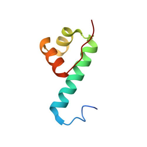
Genetic changes lead gradually to altered protein function, making deduction of the molecular basis for activity from a sequence difficult. Comparative studies provide insights into the functional consequences of specific changes. Here we present structural and biochemical studies of NtrC4, a sigma-54 activator from Aquifex aeolicus, and compare it with NtrC1 (a paralog) and NtrC (a homolog from Salmonella enterica) to provide insight into how a substantial change in regulatory mechanism may have occurred. Activity assays show that assembly of NtrC4's active oligomer is repressed by the N-terminal receiver domain, and that BeF3- addition (mimicking phosphorylation) removes this repression. Observation of assembly without activation for NtrC4 indicates that it is much less strongly repressed than NtrC1. The crystal structure of the unactivated receiver-ATPase domain combination shows a partially disrupted interface. NMR structures of the regulatory domain show that its activation mechanism is very similar to that of NtrC1. The crystal structure of the NtrC4 DNA-binding domain shows that it is dimeric and more similar in structure to NtrC than NtrC1. Electron microscope images of the ATPase-DNA-binding domain combination show formation of oligomeric rings. Sequence alignments provide insights into the distribution of activation mechanisms in this family of proteins.
- Graduate Group in Biophysics, Physical Biosciences Division, Lawrence Berkeley National Laboratory and the Department of Chemistry, University of California, Berkeley, CA 94720, USA.
Organizational Affiliation: