3E7C | pdb_00003e7c

Glucocorticoid Receptor LBD bound to GSK866


Experimental Data Snapshot

  • Method: X-RAY DIFFRACTION
  • Resolution: 2.15 Å
  • R-Value Free: 
    0.266 (Depositor), 0.217 (DCC) 
  • R-Value Work: 
    0.210 (Depositor), 0.213 (DCC) 

Starting Model: experimental
View more details

wwPDB Validation 3D Report Full Report

Validation slider image for 3E7C

Ligand Structure Quality Assessment 


This is version 1.6 of the entry. See complete history

Literature

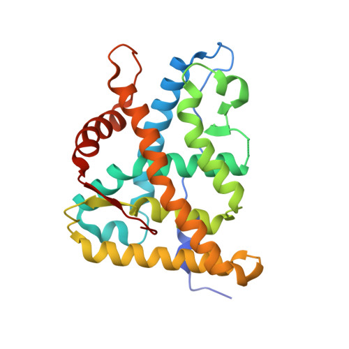
The first X-ray crystal structure of the glucocorticoid receptor bound to a non-steroidal agonist.

Madauss, K.P.Bledsoe, R.K.Mclay, I.Stewart, E.L.Uings, I.J.Weingarten, G.Williams, S.P.

(2008) Bioorg Med Chem Lett 18: 6097-6099

  • DOI: https://doi.org/10.1016/j.bmcl.2008.10.021
  • Primary Citation Related Structures: 
    3E7C

  • PubMed Abstract: 

    The amino-pyrazole 2,6-dichloro-N-ethyl benzamide 1 is a selective GR agonist with dexamethasone-like in vitro potency. Its X-ray crystal structure in the GR LBD (Glucocorticoid ligand-binding domain) is described and compared to other reported structures of steroidal GR agonists in the GR LBD (3E7C).


  • Organizational Affiliation
    • Department of Computational and Structural Chemistry, Molecular Discovery Research, GlaxoSmithKline, 5 Moore Drive, Research Triangle Park, NC 27709, USA. kevin.p.madauss@gsk.com

Macromolecule Content 

  • Total Structure Weight: 63.78 kDa 
  • Atom Count: 4,330 
  • Modeled Residue Count: 514 
  • Deposited Residue Count: 536 
  • Unique protein chains: 2

Macromolecules

Find similar proteins by:|  3D Structure
Entity ID: 1
MoleculeChains  Sequence LengthOrganismDetailsImage
Glucocorticoid receptorA,
C [auth B]
257Homo sapiensMutation(s): 2 
Gene Names: NR3C1GRL
UniProt & NIH Common Fund Data Resources
Find proteins for P04150 (Homo sapiens)
Explore P04150 
Go to UniProtKB:  P04150
PHAROS:  P04150
GTEx:  ENSG00000113580 
Entity Groups
Sequence Clusters30% Identity50% Identity70% Identity90% Identity95% Identity100% Identity
UniProt GroupP04150
Sequence Annotations
Expand
Reference Sequence
Find similar proteins by:  Sequence   |   3D Structure  
Entity ID: 2
MoleculeChains  Sequence LengthOrganismDetailsImage
Nuclear receptor coactivator 2B [auth H],
D
11Homo sapiensMutation(s): 0 
UniProt & NIH Common Fund Data Resources
Find proteins for Q15596 (Homo sapiens)
Explore Q15596 
Go to UniProtKB:  Q15596
PHAROS:  Q15596
GTEx:  ENSG00000140396 
Entity Groups
Sequence Clusters30% Identity50% Identity70% Identity90% Identity95% Identity100% Identity
UniProt GroupQ15596
Sequence Annotations
Expand
Reference Sequence

Experimental Data & Validation

Experimental Data

  • Method: X-RAY DIFFRACTION
  • Resolution: 2.15 Å
  • R-Value Free:  0.266 (Depositor), 0.217 (DCC) 
  • R-Value Work:  0.210 (Depositor), 0.213 (DCC) 
Space Group: P 61
Unit Cell:
Length ( Å )Angle ( ˚ )
a = 126.654α = 90
b = 126.654β = 90
c = 78.991γ = 120
Software Package:
Software NamePurpose
DENZOdata reduction
SCALEPACKdata scaling
PHASERphasing
PHENIXrefinement
PDB_EXTRACTdata extraction
StructureStudiodata collection
HKL-2000data reduction
HKL-2000data scaling

Structure Validation

View Full Validation Report


Ligand Structure Quality Assessment 


Entry History 

Deposition Data

Revision History  (Full details and data files)

  • Version 1.0: 2008-11-25
    Type: Initial release
  • Version 1.1: 2011-07-13
    Changes: Non-polymer description, Version format compliance
  • Version 1.2: 2014-04-02
    Changes: Source and taxonomy
  • Version 1.3: 2017-10-25
    Changes: Refinement description
  • Version 1.4: 2021-10-20
    Changes: Database references, Derived calculations
  • Version 1.5: 2024-02-21
    Changes: Data collection
  • Version 1.6: 2024-04-03
    Changes: Refinement description