3E70 | pdb_00003e70

Structures and conformations in solution of the Signal Recognition Particle Receptor from the Archaeon Pyrococcus Furiosus


Experimental Data Snapshot

  • Method: X-RAY DIFFRACTION
  • Resolution: 1.97 Å
  • R-Value Free: 
    0.263 (Depositor), 0.262 (DCC) 
  • R-Value Work: 
    0.228 (Depositor), 0.229 (DCC) 
  • R-Value Observed: 
    0.234 (Depositor) 

Starting Model: experimental
View more details

wwPDB Validation 3D Report Full Report

Validation slider image for 3E70

Ligand Structure Quality Assessment 


This is version 1.3 of the entry. See complete history

Literature

Structures of the signal recognition particle receptor from the archaeon Pyrococcus furiosus: implications for the targeting step at the membrane.

Egea, P.F.Tsuruta, H.de Leon, G.P.Napetschnig, J.Walter, P.Stroud, R.M.

(2008) PLoS One 3: e3619-e3619

  • DOI: https://doi.org/10.1371/journal.pone.0003619
  • Primary Citation Related Structures: 
    3DM9, 3DMD, 3E70

  • PubMed Abstract: 

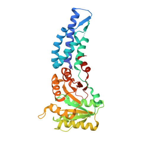
    In all organisms, a ribonucleoprotein called the signal recognition particle (SRP) and its receptor (SR) target nascent proteins from the ribosome to the translocon for secretion or membrane insertion. We present the first X-ray structures of an archeal FtsY, the receptor from the hyper-thermophile Pyrococcus furiosus (Pfu), in its free and GDP*magnesium-bound forms. The highly charged N-terminal domain of Pfu-FtsY is distinguished by a long N-terminal helix. The basic charges on the surface of this helix are likely to regulate interactions at the membrane. A peripheral GDP bound near a regulatory motif could indicate a site of interaction between the receptor and ribosomal or SRP RNAs. Small angle X-ray scattering and analytical ultracentrifugation indicate that the crystal structure of Pfu-FtsY correlates well with the average conformation in solution. Based on previous structures of two sub-complexes, we propose a model of the core of archeal and eukaryotic SRP*SR targeting complexes.


  • Organizational Affiliation
    • Department of Biochemistry and Biophysics, University of California San Francisco, San Francisco, California, USA. pascal@msg.ucsf.edu

Macromolecule Content 

  • Total Structure Weight: 37.35 kDa 
  • Atom Count: 2,484 
  • Modeled Residue Count: 307 
  • Deposited Residue Count: 328 
  • Unique protein chains: 1

Macromolecules

Find similar proteins by:|  3D Structure
Entity ID: 1
MoleculeChains  Sequence LengthOrganismDetailsImage
Signal recognition particle receptorA [auth C]328Pyrococcus furiosusMutation(s): 0 
Gene Names: PF1766
EC: 3.6.5.4
UniProt
Find proteins for Q8U051 (Pyrococcus furiosus (strain ATCC 43587 / DSM 3638 / JCM 8422 / Vc1))
Explore Q8U051 
Go to UniProtKB:  Q8U051
Entity Groups
Sequence Clusters30% Identity50% Identity70% Identity90% Identity95% Identity100% Identity
UniProt GroupQ8U051
Sequence Annotations
Expand
Reference Sequence

Experimental Data & Validation

Experimental Data

  • Method: X-RAY DIFFRACTION
  • Resolution: 1.97 Å
  • R-Value Free:  0.263 (Depositor), 0.262 (DCC) 
  • R-Value Work:  0.228 (Depositor), 0.229 (DCC) 
  • R-Value Observed: 0.234 (Depositor) 
Space Group: C 1 2 1
Unit Cell:
Length ( Å )Angle ( ˚ )
a = 113.122α = 90
b = 53.034β = 107.42
c = 61.26γ = 90
Software Package:
Software NamePurpose
PHENIXrefinement
MOSFLMdata reduction
SCALAdata scaling
PHENIXphasing

Structure Validation

View Full Validation Report


Ligand Structure Quality Assessment 


Entry History 

Deposition Data

Revision History  (Full details and data files)

  • Version 1.0: 2008-11-18
    Type: Initial release
  • Version 1.1: 2011-07-13
    Changes: Version format compliance
  • Version 1.2: 2018-01-24
    Changes: Structure summary
  • Version 1.3: 2023-08-30
    Changes: Data collection, Database references, Derived calculations, Refinement description