3E4G | pdb_00003e4g

Crystal structure of bovine coupling Factor B, G28E mutant


Experimental Data Snapshot

  • Method: X-RAY DIFFRACTION
  • Resolution: 0.96 Å
  • R-Value Free: 
    0.164 (Depositor), 0.183 (DCC) 
  • R-Value Work: 
    0.143 (Depositor) 
  • R-Value Observed: 
    0.144 (Depositor) 

Starting Model: experimental
View more details

wwPDB Validation 3D Report Full Report

Validation slider image for 3E4G

This is version 1.3 of the entry. See complete history

Literature

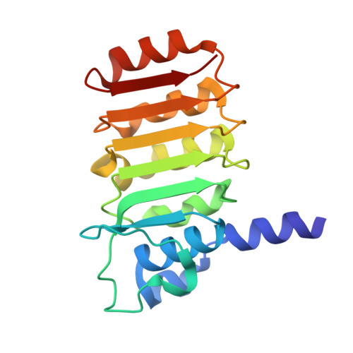
Crystal structure of bovine mitochondrial factor B at 0.96-A resolution.

Lee, J.K.Belogrudov, G.I.Stroud, R.M.

(2008) Proc Natl Acad Sci U S A 105: 13379-13384

  • DOI: https://doi.org/10.1073/pnas.0805689105
  • Primary Citation Related Structures: 
    3DZE, 3E2J, 3E3Z, 3E4G

  • PubMed Abstract: 

    Coupling factor B (FB) is a mitochondrial inner membrane polypeptide that facilitates the energy-driven catalysis of ATP synthesis in animal mitochondria by blocking a proton leak across the membrane. Here, we report the crystal structure of the bovine mitochondrial FB mutant with Gly-3-Glu substitution determined at a resolution of 0.96 A and that of the WT polypeptide at a resolution of 2.9 A. The structure reveals an oblong, oval-shaped molecule with a unique globular N-terminal domain that is proposed to be the membrane anchor domain and the capping region to the C-terminal leucine-rich repeats domain. A short N-terminal alpha-helix, which extends away from the molecule's body, is suggestive of functioning as an anchor for FB to the matrix side of the mitochondrial inner membrane. Identification of a bound Mg(2+) ion reveals that FB is a metalloprotein. We also report the cocrystal structures of FB bound with phenylarsine oxide and Cd(2+), two known inhibitors of the FB coupling activity.


  • Organizational Affiliation
    • Department of Biochemistry and Biophysics, University of California, San Francisco, CA 94158-2517, USA.

Macromolecule Content 

  • Total Structure Weight: 20.66 kDa 
  • Atom Count: 1,782 
  • Modeled Residue Count: 176 
  • Deposited Residue Count: 176 
  • Unique protein chains: 1

Macromolecules

Find similar proteins by:|  3D Structure
Entity ID: 1
MoleculeChains  Sequence LengthOrganismDetailsImage
ATP synthase subunit s, mitochondrial176Bos taurusMutation(s): 1 
Gene Names: ATP5SATPW
EC: 3.6.3.14
UniProt
Find proteins for P22027 (Bos taurus)
Explore P22027 
Go to UniProtKB:  P22027
Entity Groups
Sequence Clusters30% Identity50% Identity70% Identity90% Identity95% Identity100% Identity
UniProt GroupP22027
Sequence Annotations
Expand
Reference Sequence

Small Molecules

Ligands 2 Unique
IDChains Name / Formula / InChI Key2D Diagram3D Interactions
TRS

Query on TRS


Download:Ideal Coordinates CCD File
C [auth A]2-AMINO-2-HYDROXYMETHYL-PROPANE-1,3-DIOL
C4 H12 N O3
LENZDBCJOHFCAS-UHFFFAOYSA-O
MG

Query on MG


Download:Ideal Coordinates CCD File
B [auth A]MAGNESIUM ION
Mg
JLVVSXFLKOJNIY-UHFFFAOYSA-N

Experimental Data & Validation

Experimental Data

  • Method: X-RAY DIFFRACTION
  • Resolution: 0.96 Å
  • R-Value Free:  0.164 (Depositor), 0.183 (DCC) 
  • R-Value Work:  0.143 (Depositor) 
  • R-Value Observed: 0.144 (Depositor) 
Space Group: C 1 2 1
Unit Cell:
Length ( Å )Angle ( ˚ )
a = 107.476α = 90
b = 50.01β = 109.37
c = 36.886γ = 90
Software Package:
Software NamePurpose
MOSFLMdata reduction
SCALAdata scaling
PHASERphasing
REFMACrefinement
PDB_EXTRACTdata extraction
Blu-Icedata collection

Structure Validation

View Full Validation Report


Entry History 

Deposition Data

Revision History  (Full details and data files)

  • Version 1.0: 2008-08-26
    Type: Initial release
  • Version 1.1: 2011-07-13
    Changes: Version format compliance
  • Version 1.2: 2021-10-20
    Changes: Database references, Derived calculations
  • Version 1.3: 2023-08-30
    Changes: Data collection, Refinement description