3E0D | pdb_00003e0d

Insights into the Replisome from the Crystral Structure of the Ternary Complex of the Eubacterial DNA Polymerase III alpha-subunit


Experimental Data Snapshot

  • Method: X-RAY DIFFRACTION
  • Resolution: 4.60 Å
  • R-Value Free: 
    0.271 (Depositor) 
  • R-Value Work: 
    0.287 (Depositor), 0.300 (DCC) 

wwPDB Validation   3D Report Full Report


Ligand Structure Quality Assessment 

Created with Raphaël 2.3.0Worse 01 BetterLigand structure goodness of fit to experimental dataBest fitted DTPClick on this verticalbar to view details

This is version 1.3 of the entry. See complete history


Literature

Insights into the Replisome from the Structure of a Ternary Complex of the DNA Polymerase III alpha-Subunit.

Wing, R.A.Bailey, S.Steitz, T.A.

(2008) J Mol Biology 382: 859-869

  • DOI: https://doi.org/10.1016/j.jmb.2008.07.058
  • Primary Citation of Related Structures:  
    3E0D

  • PubMed Abstract: 

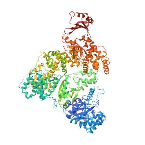
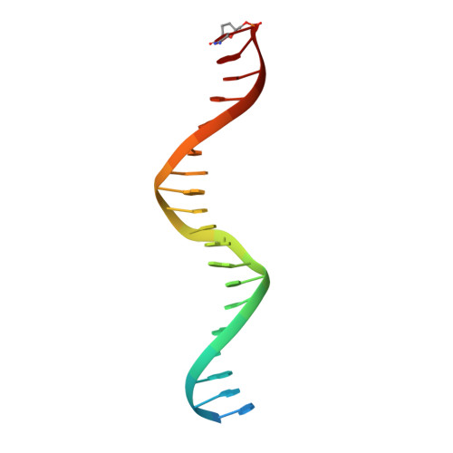
    The crystal structure of the catalytic alpha-subunit of the DNA polymerase III (Pol IIIalpha) holoenzyme bound to primer-template DNA and an incoming deoxy-nucleoside 5'-triphosphate has been determined at 4.6-A resolution. The polymerase interacts with the sugar-phosphate backbone of the DNA across its minor groove, which is made possible by significant movements of the thumb, finger, and beta-binding domains relative to their orientations in the unliganded polymerase structure. Additionally, the DNA and incoming nucleotide are bound to the active site of Pol IIIalpha nearly identically as they are in their complex with DNA polymerase beta, thereby proving that the eubacterial replicating polymerase, but not the eukaryotic replicating polymerase, is homologous to DNA polymerase beta. Finally, superimposing a recent structure of the clamp bound to DNA on this Pol IIIalpha complex with DNA places a loop of the beta-binding domain into the appropriate clamp cleft and supports a mechanism of polymerase switching.


  • Organizational Affiliation

    Department of Molecular Biophysics and Biochemistry, Yale University, New Haven, CT 06520, USA.


Macromolecules

Find similar proteins by:  (by identity cutoff)  |  3D Structure
Entity ID: 3
MoleculeChains Sequence LengthOrganismDetailsImage
DNA polymerase III subunit alphaE [auth A],
F [auth B]
1,220Thermus aquaticusMutation(s): 6 
Gene Names: dnaE
EC: 2.7.7.7
UniProt
Find proteins for Q9XDH5 (Thermus aquaticus)
Explore Q9XDH5 
Go to UniProtKB:  Q9XDH5
Entity Groups  
Sequence Clusters30% Identity50% Identity70% Identity90% Identity95% Identity100% Identity
UniProt GroupQ9XDH5
Sequence Annotations
Expand
  • Reference Sequence
Experimental Data & Validation

Experimental Data

  • Method: X-RAY DIFFRACTION
  • Resolution: 4.60 Å
  • R-Value Free:  0.271 (Depositor) 
  • R-Value Work:  0.287 (Depositor), 0.300 (DCC) 
Space Group: P 41
Unit Cell:
Length ( Å )Angle ( ˚ )
a = 149.546α = 90
b = 149.546β = 90
c = 163.338γ = 90
Software Package:
Software NamePurpose
CNSrefinement

Structure Validation

View Full Validation Report


Ligand Structure Quality Assessment 

Created with Raphaël 2.3.0Worse 01 BetterLigand structure goodness of fit to experimental dataBest fitted DTPClick on this verticalbar to view details

Entry History 

Deposition Data

Revision History  (Full details and data files)

  • Version 1.0: 2008-09-23
    Type: Initial release
  • Version 1.1: 2011-07-13
    Changes: Version format compliance
  • Version 1.2: 2021-10-20
    Changes: Data collection, Database references, Derived calculations
  • Version 1.3: 2024-02-21
    Changes: Data collection