3E0B | pdb_00003e0b

Bacillus anthracis Dihydrofolate Reductase complexed with NADPH and 2,4-diamino-5-(3-(2,5-dimethoxyphenyl)prop-1-ynyl)-6-ethylpyrimidine (UCP120B)


Experimental Data Snapshot

  • Method: X-RAY DIFFRACTION
  • Resolution: 2.25 Å
  • R-Value Free: 
    0.238 (Depositor), 0.234 (DCC) 
  • R-Value Work: 
    0.191 (Depositor), 0.191 (DCC) 
  • R-Value Observed: 
    0.193 (Depositor) 

Starting Model: experimental
View more details

wwPDB Validation 3D Report Full Report

Validation slider image for 3E0B

Ligand Structure Quality Assessment 


This is version 1.4 of the entry. See complete history

Literature

Synthetic and Crystallographic Studies of a New Inhibitor Series Targeting Bacillus anthracis Dihydrofolate Reductase

Beierlein, J.M.Frey, K.M.Bolstad, D.B.Pelphrey, P.M.Joska, T.M.Smith, A.E.Priestley, N.D.Wright, D.L.Anderson, A.C.

(2008) J Med Chem 51: 7532-7540

  • DOI: https://doi.org/10.1021/jm800776a
  • Primary Citation Related Structures: 
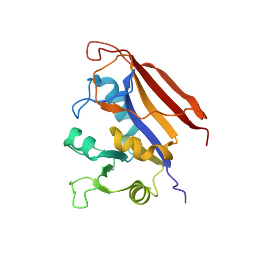
    3E0B

  • PubMed Abstract: 

    Bacillus anthracis, the causative agent of anthrax, poses a significant biodefense danger. Serious limitations in approved therapeutics and the generation of resistance have produced a compelling need for new therapeutic agents against this organism. Bacillus anthracis is known to be insensitive to the clinically used antifolate, trimethoprim, because of a lack of potency against the dihydrofolate reductase enzyme. Herein, we describe a novel lead series of B. anthracis dihydrofolate reductase inhibitors characterized by an extended trimethoprim-like scaffold. The best lead compound adds only 22 Da to the molecular weight and is 82-fold more potent than trimethoprim. An X-ray crystal structure of this lead compound bound to B. anthracis dihydrofolate reductase in the presence of NADPH was determined to 2.25 A resolution. The structure reveals several features that can be exploited for further development of this lead series.


  • Organizational Affiliation
    • Department of Pharmaceutical Sciences, University of Connecticut, 69 N. Eagleville Road, Storrs, Connecticut 06269, USA.

Macromolecule Content 

  • Total Structure Weight: 41.61 kDa 
  • Atom Count: 3,119 
  • Modeled Residue Count: 332 
  • Deposited Residue Count: 332 
  • Unique protein chains: 1

Macromolecules

Find similar proteins by:|  3D Structure
Entity ID: 1
MoleculeChains  Sequence LengthOrganismDetailsImage
Dihydrofolate reductase
A, B
166Bacillus anthracisMutation(s): 1 
Gene Names: dfrABAS2083BA_2237GBAA2237
EC: 1.5.1.3
UniProt
Find proteins for Q81R22 (Bacillus anthracis)
Explore Q81R22 
Go to UniProtKB:  Q81R22
Entity Groups
Sequence Clusters30% Identity50% Identity70% Identity90% Identity95% Identity100% Identity
UniProt GroupQ81R22
Sequence Annotations
Expand
Reference Sequence

Small Molecules

Binding Affinity Annotations 
IDSourceBinding Affinity
N22 BindingDB:  3E0B Ki: 300 (nM) from 1 assay(s)
IC50: 890 (nM) from 1 assay(s)

Experimental Data & Validation

Experimental Data

  • Method: X-RAY DIFFRACTION
  • Resolution: 2.25 Å
  • R-Value Free:  0.238 (Depositor), 0.234 (DCC) 
  • R-Value Work:  0.191 (Depositor), 0.191 (DCC) 
  • R-Value Observed: 0.193 (Depositor) 
Space Group: P 42
Unit Cell:
Length ( Å )Angle ( ˚ )
a = 78.416α = 90
b = 78.416β = 90
c = 67.092γ = 90
Software Package:
Software NamePurpose
REFMACrefinement
CBASSdata collection
HKL-2000data reduction
SCALEPACKdata scaling
PHASERphasing

Structure Validation

View Full Validation Report


Ligand Structure Quality Assessment 


Entry History 

Deposition Data

Revision History  (Full details and data files)

  • Version 1.0: 2008-11-25
    Type: Initial release
  • Version 1.1: 2011-07-13
    Changes: Version format compliance
  • Version 1.2: 2013-11-20
    Changes: Non-polymer description
  • Version 1.3: 2021-11-10
    Changes: Database references, Derived calculations
  • Version 1.4: 2023-11-01
    Changes: Data collection, Refinement description