3DX4

Golgi alpha-Mannosidase II in complex with Mannostatin analog (1R,2R,3R,4S,5R)-4-amino-5-methoxycyclopentane-1,2,3-triol


Experimental Data Snapshot

  • Method: X-RAY DIFFRACTION
  • Resolution: 1.38 Å
  • R-Value Free: 0.203 
  • R-Value Work: 0.188 
  • R-Value Observed: 0.188 

wwPDB Validation   3D Report Full Report


Ligand Structure Quality Assessment 


This is version 1.4 of the entry. See complete history


Literature

The molecular basis of inhibition of Golgi alpha-mannosidase II by mannostatin A.

Kuntz, D.A.Zhong, W.Guo, J.Rose, D.R.Boons, G.J.

(2009) Chembiochem 10: 268-277

  • DOI: https://doi.org/10.1002/cbic.200800538
  • Primary Citation of Related Structures:  
    3DX0, 3DX1, 3DX2, 3DX3, 3DX4

  • PubMed Abstract: 

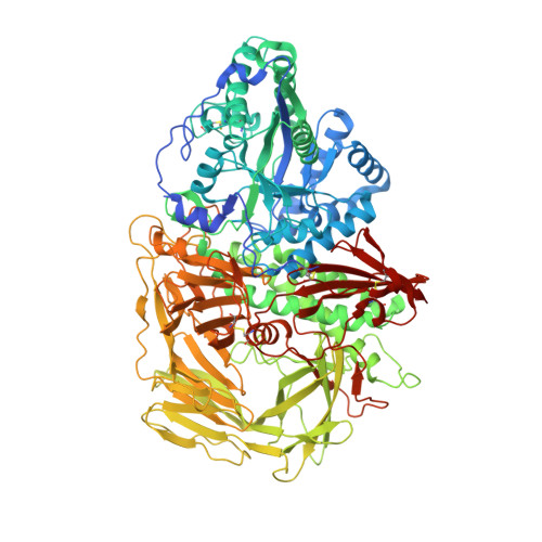
    Mannostatin A is a potent inhibitor of the mannose-trimming enzyme, Golgi alpha-mannosidase II (GMII), which acts late in the N-glycan processing pathway. Inhibition of this enzyme provides a route to blocking the transformation-associated changes in cancer cell surface oligosaccharide structures. Here, we report on the synthesis of new Mannostatin derivatives and analyze their binding in the active site of Drosophila GMII by X-ray crystallography. The results indicate that the interaction with the backbone carbonyl of Arg876 is crucial to the high potency of the inhibitor-an effect enhanced by the hydrophobic interaction between the thiomethyl group and an aromatic pocket vicinal to the cleavage site. The various structures indicate that differences in the hydration of protein-ligand complexes are also important determinants of plasticity as well as selectivity of inhibitor binding.


  • Organizational Affiliation

    Ontario Cancer Institute and Department of Medical Biophysics, University of Toronto, Toronto, Ontario M5G 1L7, Canada.


Macromolecules
Find similar proteins by:  (by identity cutoff)  |  3D Structure
Entity ID: 1
MoleculeChains Sequence LengthOrganismDetailsImage
Alpha-mannosidase 21,045Drosophila melanogasterMutation(s): 0 
Gene Names: alpha-Man-IIGmIICG18802
EC: 3.2.1.114
UniProt
Find proteins for Q24451 (Drosophila melanogaster)
Explore Q24451 
Go to UniProtKB:  Q24451
Entity Groups  
Sequence Clusters30% Identity50% Identity70% Identity90% Identity95% Identity100% Identity
UniProt GroupQ24451
Sequence Annotations
Expand
  • Reference Sequence
Small Molecules
Binding Affinity Annotations 
IDSourceBinding Affinity
GOO PDBBind:  3DX4 Ki: 76 (nM) from 1 assay(s)
Binding MOAD:  3DX4 Ki: 76 (nM) from 1 assay(s)
BindingDB:  3DX4 Ki: 76 (nM) from 1 assay(s)
Experimental Data & Validation

Experimental Data

  • Method: X-RAY DIFFRACTION
  • Resolution: 1.38 Å
  • R-Value Free: 0.203 
  • R-Value Work: 0.188 
  • R-Value Observed: 0.188 
  • Space Group: P 21 21 21
Unit Cell:
Length ( Å )Angle ( ˚ )
a = 68.769α = 90
b = 109.703β = 90
c = 138.306γ = 90
Software Package:
Software NamePurpose
REFMACrefinement
CNSrefinement
PDB_EXTRACTdata extraction
ADSCdata collection
HKL-2000data reduction
SADABSdata scaling
CNSphasing

Structure Validation

View Full Validation Report


Ligand Structure Quality Assessment 


Entry History 

Deposition Data

Revision History  (Full details and data files)

  • Version 1.0: 2009-07-07
    Type: Initial release
  • Version 1.1: 2011-07-13
    Changes: Non-polymer description, Version format compliance
  • Version 1.2: 2019-07-24
    Changes: Data collection, Derived calculations, Refinement description
  • Version 1.3: 2020-07-29
    Type: Remediation
    Reason: Carbohydrate remediation
    Changes: Advisory, Data collection, Database references, Derived calculations, Structure summary
  • Version 1.4: 2023-08-30
    Changes: Data collection, Database references, Refinement description, Structure summary