Homodimeric chicken galectin CG-1B (C-14): Crystal structure and detection of unique redox-dependent shape changes involving inter- and intrasubunit disulfide bridges by gel filtration, ultracentrifugation, site-directed mutagenesis, and peptide mass fingerprinting
Lopez-Lucendo, M.F., Solis, D., Saiz, J.L., Kaltner, H., Russwurm, R., Andre, S., Gabius, H.-J., Romero, A.(2009) J Mol Biology 386: 366-378
- PubMed: 18848566 Search on PubMed
- DOI: https://doi.org/10.1016/j.jmb.2008.09.054
- Primary Citation Related Structures:
3DUI - PubMed Abstract:
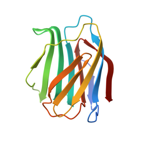
Intrafamily gene diversification has led to three prototype galectins in chicken [i.e., chicken galectin (CG)-1A, CG-1B, and CG-2] that show distinct expression profiles and developmental regulation. In order to pinpoint structural disparities among them, we determined the crystal structure of CG-1B. Alteration of the position of the Trp ring in the lectin site and the presence of only two ordered water molecules therein, as well as changes in the interface region between the two subunits, set the structure of CG-1B clearly apart from that of CG-1A. Intriguingly, the unique presence of two Cys residues at positions 2 and 7 in the N-terminal region translated into formation of an intersubunit disulfide bridge between the Cys7 residues of the homodimer in the crystal. In solution, oxidation is associated with significant shape changes in the dimeric protein and the additional occurrence of a compacted form with an intrasubunit disulfide bridge between Cys2 and Cys7. The single-site mutant C7S/C7V was not subjected to such changes, supporting the crucial role of Cys7 in redox-dependent shape changes. These results point to the functional significance of the distinctive presence of the two Cys residues in the N-terminal region of CG-1B.
- Departamento de Ciencia de Proteínas, Centro de Investigaciones Biológicas, CSIC, Madrid, Spain.
Organizational Affiliation: