3DTP

Tarantula heavy meromyosin obtained by flexible docking to Tarantula muscle thick filament Cryo-EM 3D-MAP


Experimental Data Snapshot

  • Method: ELECTRON MICROSCOPY
  • Resolution: 20.0 Å
  • Aggregation State: FILAMENT 
  • Reconstruction Method: HELICAL 

wwPDB Validation   3D Report Full Report


This is version 1.4 of the entry. See complete history


Literature

Three-Dimensional Reconstruction of Tarantula Myosin Filaments Suggests How Phosphorylation May Regulate Myosin Activity

Alamo, L.Wriggers, W.Pinto, A.Bartoli, F.Salazar, L.Zhao, F.Q.Craig, R.Padron, R.

(2008) J Mol Biol 384: 780-797

  • DOI: https://doi.org/10.1016/j.jmb.2008.10.013
  • Primary Citation of Related Structures:  
    3DTP

  • PubMed Abstract: 

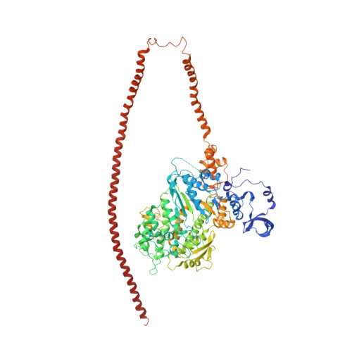
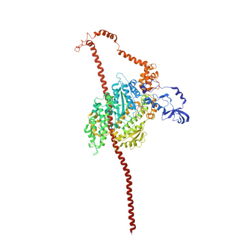
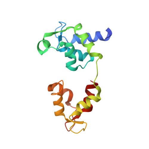
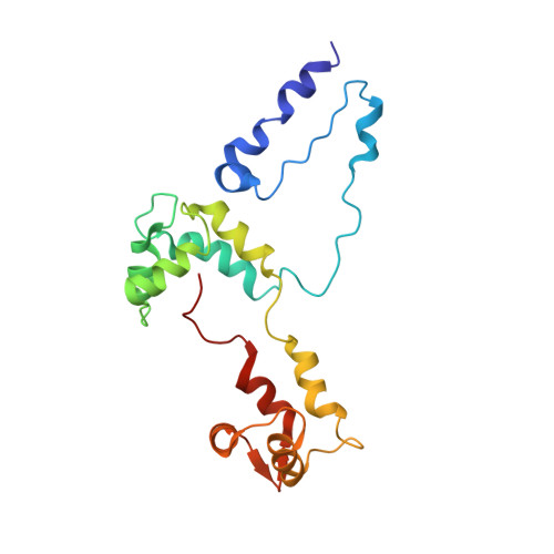
    Muscle contraction involves the interaction of the myosin heads of the thick filaments with actin subunits of the thin filaments. Relaxation occurs when this interaction is blocked by molecular switches on these filaments. In many muscles, myosin-linked regulation involves phosphorylation of the myosin regulatory light chains (RLCs). Electron microscopy of vertebrate smooth muscle myosin molecules (regulated by phosphorylation) has provided insight into the relaxed structure, revealing that myosin is switched off by intramolecular interactions between its two heads, the free head and the blocked head. Three-dimensional reconstruction of frozen-hydrated specimens revealed that this asymmetric head interaction is also present in native thick filaments of tarantula striated muscle. Our goal in this study was to elucidate the structural features of the tarantula filament involved in phosphorylation-based regulation. A new reconstruction revealed intra- and intermolecular myosin interactions in addition to those seen previously. To help interpret the interactions, we sequenced the tarantula RLC and fitted an atomic model of the myosin head that included the predicted RLC atomic structure and an S2 (subfragment 2) crystal structure to the reconstruction. The fitting suggests one intramolecular interaction, between the cardiomyopathy loop of the free head and its own S2, and two intermolecular interactions, between the cardiac loop of the free head and the essential light chain of the blocked head and between the Leu305-Gln327 interaction loop of the free head and the N-terminal fragment of the RLC of the blocked head. These interactions, added to those previously described, would help switch off the thick filament. Molecular dynamics simulations suggest how phosphorylation could increase the helical content of the RLC N-terminus, weakening these interactions, thus releasing both heads and activating the thick filament.


  • Organizational Affiliation

    Departamento de Biología Estructural, Instituto Venezolano de Investigaciones Científicas, Caracas, Venezuela.


Macromolecules
Find similar proteins by:  (by identity cutoff)  |  3D Structure
Entity ID: 1
MoleculeChains Sequence LengthOrganismDetailsImage
Myosin-11,Myosin-7971Gallus gallusHomo sapiens
This entity is chimeric
Mutation(s): 0 
Gene Names: MYH11MYH7MYHCB
UniProt & NIH Common Fund Data Resources
Find proteins for P10587 (Gallus gallus)
Explore P10587 
Go to UniProtKB:  P10587
Find proteins for P12883 (Homo sapiens)
Explore P12883 
Go to UniProtKB:  P12883
PHAROS:  P12883
GTEx:  ENSG00000092054 
Entity Groups  
Sequence Clusters30% Identity50% Identity70% Identity90% Identity95% Identity100% Identity
UniProt GroupsP12883P10587
Sequence Annotations
Expand
  • Reference Sequence
Find similar proteins by:  (by identity cutoff)  |  3D Structure
Entity ID: 2
MoleculeChains Sequence LengthOrganismDetailsImage
Myosin-11,Myosin-7973Gallus gallusHomo sapiens
This entity is chimeric
Mutation(s): 0 
Gene Names: MYH11MYH7MYHCB
UniProt & NIH Common Fund Data Resources
Find proteins for P10587 (Gallus gallus)
Explore P10587 
Go to UniProtKB:  P10587
Find proteins for P12883 (Homo sapiens)
Explore P12883 
Go to UniProtKB:  P12883
PHAROS:  P12883
GTEx:  ENSG00000092054 
Entity Groups  
Sequence Clusters30% Identity50% Identity70% Identity90% Identity95% Identity100% Identity
UniProt GroupsP12883P10587
Sequence Annotations
Expand
  • Reference Sequence
Find similar proteins by:  (by identity cutoff)  |  3D Structure
Entity ID: 3
MoleculeChains Sequence LengthOrganismDetailsImage
Myosin light polypeptide 6
C, D
150Gallus gallusMutation(s): 0 
Gene Names: MYL6
UniProt
Find proteins for P02607 (Gallus gallus)
Explore P02607 
Go to UniProtKB:  P02607
Entity Groups  
Sequence Clusters30% Identity50% Identity70% Identity90% Identity95% Identity100% Identity
UniProt GroupP02607
Sequence Annotations
Expand
  • Reference Sequence
Find similar proteins by:  (by identity cutoff)  |  3D Structure
Entity ID: 4
MoleculeChains Sequence LengthOrganismDetailsImage
Myosin II regulatory light chain
E, F
196Avicularia aviculariaMutation(s): 0 
UniProt
Find proteins for B4XT43 (Avicularia avicularia)
Explore B4XT43 
Go to UniProtKB:  B4XT43
Entity Groups  
Sequence Clusters30% Identity50% Identity70% Identity90% Identity95% Identity100% Identity
UniProt GroupB4XT43
Sequence Annotations
Expand
  • Reference Sequence
Experimental Data & Validation

Experimental Data

  • Method: ELECTRON MICROSCOPY
  • Resolution: 20.0 Å
  • Aggregation State: FILAMENT 
  • Reconstruction Method: HELICAL 

Structure Validation

View Full Validation Report


Entry History 

Deposition Data

Revision History  (Full details and data files)

  • Version 1.0: 2008-10-07
    Type: Initial release
  • Version 1.1: 2011-07-13
    Changes: Version format compliance
  • Version 1.2: 2015-07-22
    Changes: Source and taxonomy
  • Version 1.3: 2016-12-14
    Changes: Other
  • Version 1.4: 2020-01-29
    Changes: Author supporting evidence, Data collection, Database references, Source and taxonomy, Structure summary