3DR4 | pdb_00003dr4

GDP-perosamine synthase K186A mutant from Caulobacter crescentus with bound sugar ligand


Experimental Data Snapshot

  • Method: X-RAY DIFFRACTION
  • Resolution: 1.60 Å
  • R-Value Free: 
    0.238 (Depositor) 
  • R-Value Work: 
    0.166 (Depositor), 0.170 (DCC) 

Starting Model: experimental
View more details

wwPDB Validation   3D Report Full Report


Ligand Structure Quality Assessment 

Created with Raphaël 2.3.0Worse 01 BetterLigand structure goodness of fit to experimental dataBest fitted G4MClick on this verticalbar to view details

This is version 1.4 of the entry. See complete history


Literature

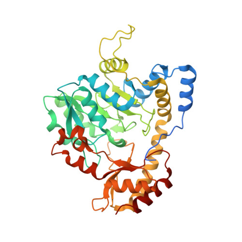
Accommodation of GDP-linked sugars in the active site of GDP-perosamine synthase

Cook, P.D.Carney, A.E.Holden, H.M.

(2008) Biochemistry 47: 10685-10693

  • DOI: https://doi.org/10.1021/bi801309q
  • Primary Citation of Related Structures:  
    3DR4, 3DR7

  • PubMed Abstract: 

    Perosamine (4-amino-4,6-dideoxy- d-mannose), or its N-acetylated form, is one of several dideoxy sugars found in the O-antigens of such infamous Gram-negative bacteria as Vibrio cholerae O1 and Escherichia coli O157:H7. It is added to the bacterial O-antigen via a nucleotide-linked version, namely GDP-perosamine. Three enzymes are required for the biosynthesis of GDP-perosamine starting from mannose 1-phosphate. The focus of this investigation is GDP-perosamine synthase from Caulobacter crescentus, which catalyzes the final step in GDP-perosamine synthesis, the conversion of GDP-4-keto-6-deoxymannose to GDP-perosamine. The enzyme is PLP-dependent and belongs to the aspartate aminotransferase superfamily. It contains the typically conserved active site lysine residue, which forms a Schiff base with the PLP cofactor. Two crystal structures were determined for this investigation: a site-directed mutant protein (K186A) complexed with GDP-perosamine and the wild-type enzyme complexed with an unnatural ligand, GDP-3-deoxyperosamine. These structures, determined to 1.6 and 1.7 A resolution, respectively, revealed the manner in which products, and presumably substrates, are accommodated within the active site pocket of GDP-perosamine synthase. Additional kinetic analyses using both the natural and unnatural substrates revealed that the K m for the unnatural substrate was unperturbed relative to that of the natural substrate, but the k cat was lowered by a factor of approximately 200. Taken together, these studies shed light on why GDP-perosamine synthase functions as an aminotransferase whereas another very similar PLP-dependent enzyme, GDP-4-keto-6-deoxy- d-mannose 3-dehydratase or ColD, catalyzes a dehydration reaction using the same substrate.


  • Organizational Affiliation

    Department of Biochemistry, University of Wisconsin, Madison, Wisconsin 53706, USA.


Macromolecules
Find similar proteins by:  (by identity cutoff)  |  3D Structure
Entity ID: 1
MoleculeChains Sequence LengthOrganismDetailsImage
Putative perosamine synthetase
A, B, C, D
391Caulobacter vibrioidesMutation(s): 1 
Gene Names: per
EC: 2.6.1.102
UniProt
Find proteins for Q9A9H3 (Caulobacter vibrioides (strain ATCC 19089 / CIP 103742 / CB 15))
Explore Q9A9H3 
Go to UniProtKB:  Q9A9H3
Entity Groups  
Sequence Clusters30% Identity50% Identity70% Identity90% Identity95% Identity100% Identity
UniProt GroupQ9A9H3
Sequence Annotations
Expand
  • Reference Sequence
Small Molecules
Ligands 2 Unique
IDChains Name / Formula / InChI Key2D Diagram3D Interactions
G4M
Query on G4M

Download Ideal Coordinates CCD File 
E [auth A],
F [auth B],
I [auth C],
J [auth D]
[(2R,3S,4R,5R)-5-(2-amino-6-oxo-1,6-dihydro-9H-purin-9-yl)-3,4-dihydroxytetrahydrofuran-2-yl]methyl (2R,3S,4S,5S,6R)-3,4-dihydroxy-5-[({3-hydroxy-2-methyl-5-[(phosphonooxy)methyl]pyridin-4-yl}methyl)amino]-6-methyltetrahydro-2H-pyran-2-yl dihydrogen diphosphate
C24 H36 N7 O19 P3
BNJJFFPICXKOFM-VMLLIFSYSA-N
EDO
Query on EDO

Download Ideal Coordinates CCD File 
G [auth B],
H [auth B]
1,2-ETHANEDIOL
C2 H6 O2
LYCAIKOWRPUZTN-UHFFFAOYSA-N
Experimental Data & Validation

Experimental Data

  • Method: X-RAY DIFFRACTION
  • Resolution: 1.60 Å
  • R-Value Free:  0.238 (Depositor) 
  • R-Value Work:  0.166 (Depositor), 0.170 (DCC) 
Space Group: P 1 21 1
Unit Cell:
Length ( Å )Angle ( ˚ )
a = 50.099α = 90
b = 151.921β = 102.09
c = 105.747γ = 90
Software Package:
Software NamePurpose
EPMRphasing
TNTrefinement
HKL-2000data reduction
SCALEPACKdata scaling

Structure Validation

View Full Validation Report


Ligand Structure Quality Assessment 

Created with Raphaël 2.3.0Worse 01 BetterLigand structure goodness of fit to experimental dataBest fitted G4MClick on this verticalbar to view details

Entry History 

Deposition Data

Revision History  (Full details and data files)

  • Version 1.0: 2008-10-14
    Type: Initial release
  • Version 1.1: 2011-07-13
    Changes: Version format compliance
  • Version 1.2: 2021-07-28
    Changes: Database references, Derived calculations, Refinement description, Source and taxonomy
  • Version 1.3: 2021-10-20
    Changes: Database references
  • Version 1.4: 2023-08-30
    Changes: Data collection, Refinement description