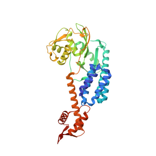
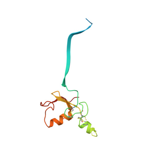
3DPL

Structural Insights into NEDD8 Activation of Cullin-RING Ligases: Conformational Control of Conjugation.


Experimental Data Snapshot

  • Method: X-RAY DIFFRACTION
  • Resolution: 2.60 Å
  • R-Value Free: 0.277 
  • R-Value Work: 0.244 
  • R-Value Observed: 0.244 

wwPDB Validation   3D Report Full Report


This is version 1.3 of the entry. See complete history


Literature

Structural insights into NEDD8 activation of cullin-RING ligases: conformational control of conjugation.

Duda, D.M.Borg, L.A.Scott, D.C.Hunt, H.W.Hammel, M.Schulman, B.A.

(2008) Cell 134: 995-1006

  • DOI: https://doi.org/10.1016/j.cell.2008.07.022
  • Primary Citation of Related Structures:  
    3DPL, 3DQV

  • PubMed Abstract: 

    Cullin-RING ligases (CRLs) comprise the largest ubiquitin E3 subclass, in which a central cullin subunit links a substrate-binding adaptor with an E2-binding RING. Covalent attachment of the ubiquitin-like protein NEDD8 to a conserved C-terminal domain (ctd) lysine stimulates CRL ubiquitination activity and prevents binding of the inhibitor CAND1. Here we report striking conformational rearrangements in the crystal structure of NEDD8~Cul5(ctd)-Rbx1 and SAXS analysis of NEDD8~Cul1(ctd)-Rbx1 relative to their unmodified counterparts. In NEDD8ylated CRL structures, the cullin WHB and Rbx1 RING subdomains are dramatically reoriented, eliminating a CAND1-binding site and imparting multiple potential catalytic geometries to an associated E2. Biochemical analyses indicate that the structural malleability is important for both CRL NEDD8ylation and subsequent ubiquitination activities. Thus, our results point to a conformational control of CRL activity, with ligation of NEDD8 shifting equilibria to disfavor inactive CAND1-bound closed architectures, and favor dynamic, open forms that promote polyubiquitination.


  • Organizational Affiliation

    Howard Hughes Medical Institute, St Jude Children's Research Hospital, Memphis, TN 38105, USA.


Macromolecules
Find similar proteins by:  (by identity cutoff)  |  3D Structure
Entity ID: 1
MoleculeChains Sequence LengthOrganismDetailsImage
Cullin-5A [auth C]382Homo sapiensMutation(s): 3 
Gene Names: CUL5VACM1
UniProt & NIH Common Fund Data Resources
Find proteins for Q93034 (Homo sapiens)
Explore Q93034 
Go to UniProtKB:  Q93034
PHAROS:  Q93034
GTEx:  ENSG00000166266 
Entity Groups  
Sequence Clusters30% Identity50% Identity70% Identity90% Identity95% Identity100% Identity
UniProt GroupQ93034
Sequence Annotations
Expand
  • Reference Sequence
Find similar proteins by:  (by identity cutoff)  |  3D Structure
Entity ID: 2
MoleculeChains Sequence LengthOrganismDetailsImage
RING-box protein 1B [auth R]106Homo sapiensMutation(s): 0 
Gene Names: RBX1RNF75ROC1
UniProt & NIH Common Fund Data Resources
Find proteins for P62877 (Homo sapiens)
Explore P62877 
Go to UniProtKB:  P62877
PHAROS:  P62877
GTEx:  ENSG00000100387 
Entity Groups  
Sequence Clusters30% Identity50% Identity70% Identity90% Identity95% Identity100% Identity
UniProt GroupP62877
Sequence Annotations
Expand
  • Reference Sequence
Experimental Data & Validation

Experimental Data

  • Method: X-RAY DIFFRACTION
  • Resolution: 2.60 Å
  • R-Value Free: 0.277 
  • R-Value Work: 0.244 
  • R-Value Observed: 0.244 
  • Space Group: P 21 21 21
Unit Cell:
Length ( Å )Angle ( ˚ )
a = 62.965α = 90
b = 65.51β = 90
c = 141.116γ = 90
Software Package:
Software NamePurpose
ADSCdata collection
CNSrefinement
HKL-2000data reduction
HKL-2000data scaling
CNSphasing

Structure Validation

View Full Validation Report


Entry History 

Deposition Data

Revision History  (Full details and data files)

  • Version 1.0: 2008-09-30
    Type: Initial release
  • Version 1.1: 2011-07-13
    Changes: Version format compliance
  • Version 1.2: 2021-10-20
    Changes: Database references, Derived calculations
  • Version 1.3: 2024-10-09
    Changes: Data collection, Structure summary