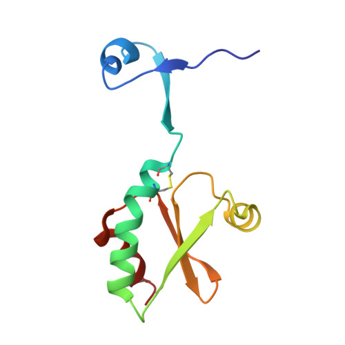
3DIE

Domain swapping of Staphylococcus Aureus thioredoxin W28A mutant


Experimental Data Snapshot

  • Method: X-RAY DIFFRACTION
  • Resolution: 1.85 Å
  • R-Value Free: 0.220 
  • R-Value Work: 0.192 
  • R-Value Observed: 0.193 

wwPDB Validation   3D Report Full Report


This is version 1.3 of the entry. See complete history


Literature

Coupling of domain swapping to kinetic stability in a thioredoxin mutant

Garcia-Pino, A.Martinez-Rodriguez, S.Wahni, K.Wyns, L.Loris, R.Messens, J.

(2009) J Mol Biol 385: 1590-1599

  • DOI: https://doi.org/10.1016/j.jmb.2008.11.040
  • Primary Citation of Related Structures:  
    3DIE

  • PubMed Abstract: 

    The thioredoxin (Trx) fold is a small monomeric domain that is ubiquitous in redox-active enzymes. Trxs are characterized by a typical WCGPC active-site sequence motif. A single active-site mutation of the tryptophan to an alanine in Staphylococcus aureus Trx converts the oxidized protein into a biologically inactive domain-swapped dimer. While the monomeric protein unfolds reversibly in a two-state manner, the oxidized dimeric form is kinetically stable and converts to the monomeric form upon refolding. After reduction, the half-life of the dimer decreases many orders of magnitude to approximately 4.3 h, indicating that the active-site disulfide between Cys29 and Cys32 is an important determinant for the kinetics of unfolding. We propose kinetic stability as a possible evolutionary strategy in the evolution of multimeric proteins from their monomeric ancestors by domain swapping, which, for this biologically inactive Trx mutant, turned out to be an evolutionary dead end.


  • Organizational Affiliation

    Department of Molecular and Cellular Interactions, Vlaams Instituut voor Biotechnologie, Brussels, Belgium.


Macromolecules
Find similar proteins by:  (by identity cutoff)  |  3D Structure
Entity ID: 1
MoleculeChains Sequence LengthOrganismDetailsImage
Thioredoxin
A, B
106Staphylococcus aureusMutation(s): 1 
Gene Names: trxA
UniProt
Find proteins for P0A0K6 (Staphylococcus aureus)
Explore P0A0K6 
Go to UniProtKB:  P0A0K6
Entity Groups  
Sequence Clusters30% Identity50% Identity70% Identity90% Identity95% Identity100% Identity
UniProt GroupP0A0K6
Sequence Annotations
Expand
  • Reference Sequence
Experimental Data & Validation

Experimental Data

  • Method: X-RAY DIFFRACTION
  • Resolution: 1.85 Å
  • R-Value Free: 0.220 
  • R-Value Work: 0.192 
  • R-Value Observed: 0.193 
  • Space Group: P 21 21 2
Unit Cell:
Length ( Å )Angle ( ˚ )
a = 88.703α = 90
b = 84.451β = 90
c = 34.699γ = 90
Software Package:
Software NamePurpose
REFMACrefinement
PDB_EXTRACTdata extraction
HKL-2000data collection
DENZOdata reduction
SCALEPACKdata scaling
PHASERphasing

Structure Validation

View Full Validation Report


Entry History 

Deposition Data

Revision History  (Full details and data files)

  • Version 1.0: 2009-03-24
    Type: Initial release
  • Version 1.1: 2011-07-13
    Changes: Version format compliance
  • Version 1.2: 2021-11-10
    Changes: Data collection, Database references, Derived calculations
  • Version 1.3: 2024-10-30
    Changes: Data collection, Structure summary