3DIB | pdb_00003dib

Crystal structure of bovine pancreatic ribonuclease A variant (I106A)


Experimental Data Snapshot

  • Method: X-RAY DIFFRACTION
  • Resolution: 1.40 Å
  • R-Value Free: 
    0.218 (Depositor), 0.219 (DCC) 
  • R-Value Work: 
    0.185 (Depositor), 0.185 (DCC) 
  • R-Value Observed: 
    0.185 (Depositor) 

Starting Model: experimental
View more details

wwPDB Validation 3D Report Full Report

Validation slider image for 3DIB

This is version 1.5 of the entry. See complete history

Literature

X-ray crystallographic studies of RNase A variants engineered at the most destabilizing positions of the main hydrophobic core: further insight into protein stability

Kurpiewska, K.Font, J.Ribo, M.Vilanova, M.Lewinski, K.

(2009) Proteins 77: 658-669

  • DOI: https://doi.org/10.1002/prot.22480
  • Primary Citation Related Structures: 
    3DH5, 3DH6, 3DI7, 3DI8, 3DI9, 3DIB, 3DIC

  • PubMed Abstract: 

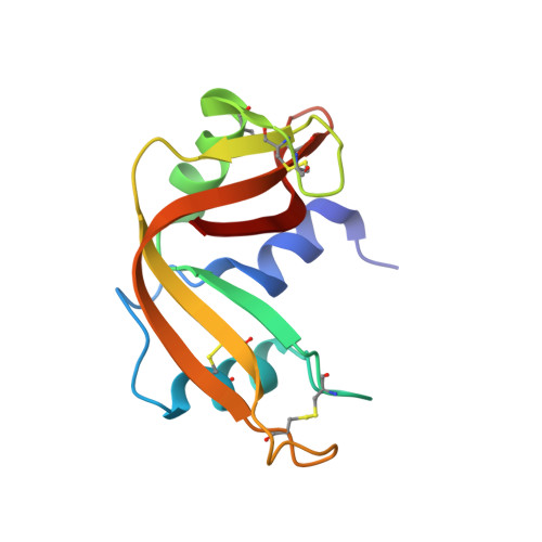
    To investigate the structural origin of decreased pressure and temperature stability, the crystal structure of bovine pancreatic ribonuclease A variants V47A, V54A, V57A, I81A, I106A, and V108A was solved at 1.4-2.0 A resolution and compared with the structure of wild-type protein. The introduced mutations had only minor influence on the global structure of ribonuclease A. The structural changes had individual character that depends on the localization of mutated residue, however, they seemed to expand from mutation site to the rest of the structure. Several different parameters have been evaluated to find correlation with decrease of free energy of unfolding DeltaDeltaG(T), and the most significant correlation was found for main cavity volume change. Analysis of the difference distance matrices revealed that the ribonuclease A molecule is organized into five relatively rigid subdomains with individual response to mutation. This behavior consistent with results of unfolding experiments is an intrinsic feature of ribonuclease A that might be surviving remnants of folding intermediates and reflects the dynamic nature of the molecule.


  • Organizational Affiliation
    • Faculty of Chemistry, Jagiellonian University, Ingardena 3, Kraków 30-060, Poland.

Macromolecule Content 

  • Total Structure Weight: 13.94 kDa 
  • Atom Count: 1,197 
  • Modeled Residue Count: 124 
  • Deposited Residue Count: 124 
  • Unique protein chains: 1

Macromolecules

Find similar proteins by:|  3D Structure
Entity ID: 1
MoleculeChains  Sequence LengthOrganismDetailsImage
Ribonuclease pancreatic124Bos taurusMutation(s): 1 
Gene Names: RNASE1RNS1
EC: 3.1.27.5 (PDB Primary Data), 4.6.1.18 (UniProt)
UniProt
Find proteins for P61823 (Bos taurus)
Explore P61823 
Go to UniProtKB:  P61823
Entity Groups
Sequence Clusters30% Identity50% Identity70% Identity90% Identity95% Identity100% Identity
UniProt GroupP61823
Sequence Annotations
Expand
Reference Sequence

Experimental Data & Validation

Experimental Data

  • Method: X-RAY DIFFRACTION
  • Resolution: 1.40 Å
  • R-Value Free:  0.218 (Depositor), 0.219 (DCC) 
  • R-Value Work:  0.185 (Depositor), 0.185 (DCC) 
  • R-Value Observed: 0.185 (Depositor) 
Space Group: P 32 2 1
Unit Cell:
Length ( Å )Angle ( ˚ )
a = 64.331α = 90
b = 64.331β = 90
c = 63.922γ = 120
Software Package:
Software NamePurpose
DENZOdata reduction
SCALEPACKdata scaling
CNSrefinement
PDB_EXTRACTdata extraction
HKL-2000data collection
CNSphasing

Structure Validation

View Full Validation Report


Entry History 

Deposition Data

Revision History  (Full details and data files)

  • Version 1.0: 2008-07-15
    Type: Initial release
  • Version 1.1: 2011-07-13
    Changes: Derived calculations, Version format compliance
  • Version 1.2: 2017-10-25
    Changes: Refinement description
  • Version 1.3: 2021-11-10
    Changes: Database references, Derived calculations
  • Version 1.4: 2023-11-01
    Changes: Data collection, Refinement description
  • Version 1.5: 2024-10-30
    Changes: Structure summary