3DI6 | pdb_00003di6

HIV-1 RT with pyridazinone non-nucleoside inhibitor


Experimental Data Snapshot

  • Method: X-RAY DIFFRACTION
  • Resolution: 2.65 Å
  • R-Value Free: 
    0.265 (Depositor), 0.266 (DCC) 
  • R-Value Work: 
    0.217 (Depositor), 0.222 (DCC) 

wwPDB Validation 3D Report Full Report

Validation slider image for 3DI6

Ligand Structure Quality Assessment 


This is version 1.2 of the entry. See complete history

Literature

Discovery and optimization of pyridazinone non-nucleoside inhibitors of HIV-1 reverse transcriptase.

Sweeney, Z.K.Dunn, J.P.Li, Y.Heilek, G.Dunten, P.Elworthy, T.R.Han, X.Harris, S.F.Hirschfeld, D.R.Hogg, J.H.Huber, W.Kaiser, A.C.Kertesz, D.J.Kim, W.Mirzadegan, T.Roepel, M.G.Saito, Y.D.Silva, T.M.Swallow, S.Tracy, J.L.Villasenor, A.Vora, H.Zhou, A.S.Klumpp, K.

(2008) Bioorg Med Chem Lett 18: 4352-4354

  • DOI: https://doi.org/10.1016/j.bmcl.2008.06.072
  • Primary Citation Related Structures: 
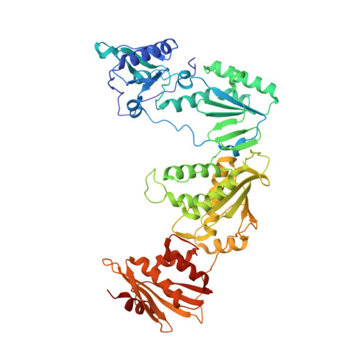
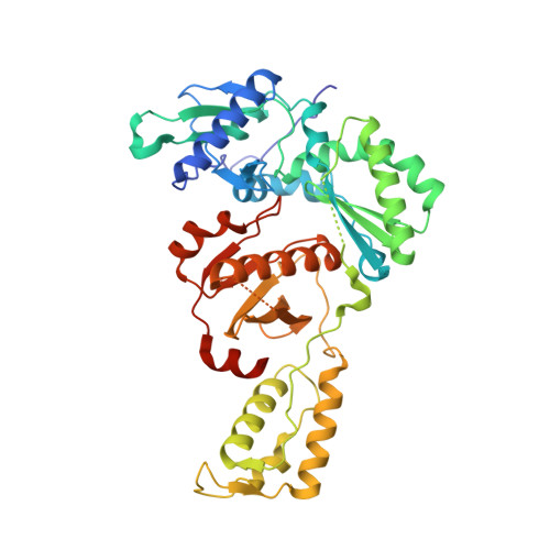
    3DI6

  • PubMed Abstract: 

    A series of benzyl pyridazinones were evaluated as HIV-1 non-nucleoside reverse transcriptase inhibitors (NNRTIs). Several members of this series showed good activity against the wild-type virus and NNRTI-resistant viruses. The binding of inhibitor 5a to HIV-RT was analyzed by surface plasmon resonance spectroscopy. Pharmacokinetic studies of 5a in rat and dog demonstrated that this compound has good oral bioavailability in animal species. The crystal structure of a complex between HIV-RT and inhibitor 4c is also described.


  • Organizational Affiliation
    • Roche Palo Alto LLC, 3431 Hillview Avenue, Palo Alto, CA 94304, USA. zachary.sweeney@roche.com

Macromolecule Content 

  • Total Structure Weight: 116.44 kDa 
  • Atom Count: 7,908 
  • Modeled Residue Count: 951 
  • Deposited Residue Count: 1,001 
  • Unique protein chains: 2

Macromolecules

Find similar proteins by:|  3D Structure
Entity ID: 1
MoleculeChains  Sequence LengthOrganismDetailsImage
Reverse transcriptase/ribonuclease H561HIV-1 M:B_HXB2RMutation(s): 0 
Gene Names: gag-pol
EC: 2.7.7.49 (PDB Primary Data), 2.7.7.7 (PDB Primary Data), 3.1.26.4 (PDB Primary Data)
UniProt
Find proteins for P04585 (Human immunodeficiency virus type 1 group M subtype B (isolate HXB2))
Explore P04585 
Go to UniProtKB:  P04585
Entity Groups
Sequence Clusters30% Identity50% Identity70% Identity90% Identity95% Identity100% Identity
UniProt GroupP04585
Sequence Annotations
Expand
Reference Sequence
Find similar proteins by:|  3D Structure
Entity ID: 2
MoleculeChains  Sequence LengthOrganismDetailsImage
p51 RT440HIV-1 M:B_HXB2RMutation(s): 0 
Gene Names: gag-pol
UniProt
Find proteins for P04585 (Human immunodeficiency virus type 1 group M subtype B (isolate HXB2))
Explore P04585 
Go to UniProtKB:  P04585
Entity Groups
Sequence Clusters30% Identity50% Identity70% Identity90% Identity95% Identity100% Identity
UniProt GroupP04585
Sequence Annotations
Expand
Reference Sequence

Small Molecules

Ligands 1 Unique
IDChains Name / Formula / InChI Key2D Diagram3D Interactions
PDZ

Query on PDZ


Download:Ideal Coordinates CCD File
C [auth A]6-(4-chloro-2-fluoro-3-phenoxybenzyl)pyridazin-3(2H)-one
C17 H12 Cl F N2 O2
NOVPOXGMADEKPP-UHFFFAOYSA-N

Experimental Data & Validation

Experimental Data

  • Method: X-RAY DIFFRACTION
  • Resolution: 2.65 Å
  • R-Value Free:  0.265 (Depositor), 0.266 (DCC) 
  • R-Value Work:  0.217 (Depositor), 0.222 (DCC) 
Space Group: C 2 2 21
Unit Cell:
Length ( Å )Angle ( ˚ )
a = 117.046α = 90
b = 153.51β = 90
c = 154.551γ = 90
Software Package:
Software NamePurpose
PHASERphasing
REFMACrefinement
HKL-2000data reduction
HKL-2000data scaling

Structure Validation

View Full Validation Report


Ligand Structure Quality Assessment 


Entry History 

Deposition Data

Revision History  (Full details and data files)

  • Version 1.0: 2008-08-19
    Type: Initial release
  • Version 1.1: 2011-07-13
    Changes: Version format compliance
  • Version 1.2: 2024-02-21
    Changes: Data collection, Database references, Derived calculations