3DHB | pdb_00003dhb

1.4 Angstrom Structure of N-Acyl Homoserine Lactone Hydrolase with the Product N-Hexanoyl-L-Homoserine Bound at The Catalytic Metal Center


Experimental Data Snapshot

  • Method: X-RAY DIFFRACTION
  • Resolution: 1.40 Å
  • R-Value Free: 
    0.184 (Depositor), 0.181 (DCC) 
  • R-Value Work: 
    0.142 (Depositor), 0.137 (DCC) 
  • R-Value Observed: 
    0.144 (Depositor) 

Starting Model: experimental
View more details

wwPDB Validation 3D Report Full Report

Validation slider image for 3DHB

Ligand Structure Quality Assessment 


This is version 1.3 of the entry. See complete history

Literature

Mechanism of the quorum-quenching lactonase (AiiA) from Bacillus thuringiensis. 1. Product-bound structures.

Liu, D.Momb, J.Thomas, P.W.Moulin, A.Petsko, G.A.Fast, W.Ringe, D.

(2008) Biochemistry 47: 7706-7714

  • DOI: https://doi.org/10.1021/bi800368y
  • Primary Citation Related Structures: 
    3DHA, 3DHB, 3DHC

  • PubMed Abstract: 

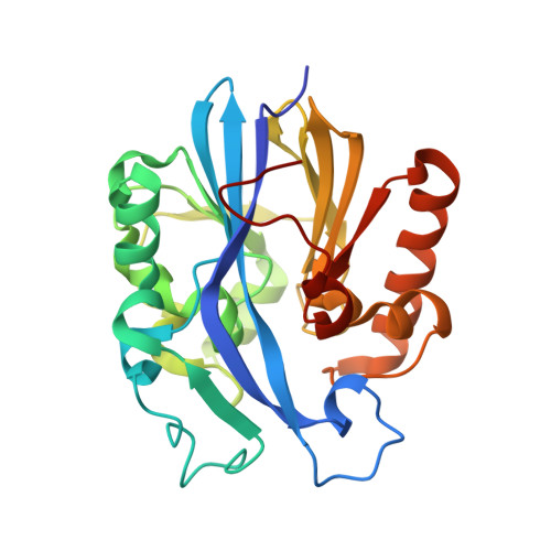
    Enzymes capable of hydrolyzing N-acyl- l-homoserine lactones (AHLs) used in some bacterial quorum-sensing pathways are of considerable interest for their ability to block undesirable phenotypes. Most known AHL hydrolases that catalyze ring opening (AHL lactonases) are members of the metallo-beta-lactamase enzyme superfamily and rely on a dinuclear zinc site for catalysis and stability. Here we report the three-dimensional structures of three product complexes formed with the AHL lactonase from Bacillus thuringiensis. Structures of the lactonase bound with two different concentrations of the ring-opened product of N-hexanoyl- l-homoserine lactone are determined at 0.95 and 1.4 A resolution and exhibit different product configurations. A structure of the ring-opened product of the non-natural N-hexanoyl- l-homocysteine thiolactone at 1.3 A resolution is also determined. On the basis of these product-bound structures, a substrate-binding model is presented that differs from previous proposals. Additionally, the proximity of the product to active-site residues and observed changes in protein conformation and metal coordination provide insight into the catalytic mechanism of this quorum-quenching metalloenzyme.


  • Organizational Affiliation
    • Department of Chemistry, Brandeis University, Waltham, Massachusetts 02454-9110, USA.

Macromolecule Content 

  • Total Structure Weight: 29.66 kDa 
  • Atom Count: 2,440 
  • Modeled Residue Count: 250 
  • Deposited Residue Count: 254 
  • Unique protein chains: 1

Macromolecules

Find similar proteins by:|  3D Structure
Entity ID: 1
MoleculeChains  Sequence LengthOrganismDetailsImage
N-Acyl Homoserine Lactone Hydrolase254Bacillus thuringiensis serovar kurstakiMutation(s): 0 
Gene Names: aiiA
EC: 3.1.1 (PDB Primary Data), 3.1.1.81 (UniProt)
UniProt
Find proteins for P0CJ63 (Bacillus thuringiensis subsp. kurstaki)
Explore P0CJ63 
Go to UniProtKB:  P0CJ63
Entity Groups
Sequence Clusters30% Identity50% Identity70% Identity90% Identity95% Identity100% Identity
UniProt GroupP0CJ63
Sequence Annotations
Expand
Reference Sequence

Small Molecules

Ligands 3 Unique
IDChains Name / Formula / InChI Key2D Diagram3D Interactions
C6L

Query on C6L


Download:Ideal Coordinates CCD File
D [auth A]N-hexanoyl-L-homoserine
C10 H19 N O4
BJLILZXVIKTHKQ-QMMMGPOBSA-N
GOL

Query on GOL


Download:Ideal Coordinates CCD File
E [auth A]
F [auth A]
G [auth A]
H [auth A]
I [auth A]
E [auth A],
F [auth A],
G [auth A],
H [auth A],
I [auth A],
J [auth A],
K [auth A]
GLYCEROL
C3 H8 O3
PEDCQBHIVMGVHV-UHFFFAOYSA-N
ZN

Query on ZN


Download:Ideal Coordinates CCD File
B [auth A],
C [auth A]
ZINC ION
Zn
PTFCDOFLOPIGGS-UHFFFAOYSA-N

Experimental Data & Validation

Experimental Data

  • Method: X-RAY DIFFRACTION
  • Resolution: 1.40 Å
  • R-Value Free:  0.184 (Depositor), 0.181 (DCC) 
  • R-Value Work:  0.142 (Depositor), 0.137 (DCC) 
  • R-Value Observed: 0.144 (Depositor) 
Space Group: P 21 21 21
Unit Cell:
Length ( Å )Angle ( ˚ )
a = 55.438α = 90
b = 55.583β = 90
c = 79.866γ = 90
Software Package:
Software NamePurpose
REFMACrefinement
Blu-Icedata collection
HKL-2000data reduction
HKL-2000data scaling
PHASERphasing

Structure Validation

View Full Validation Report


Ligand Structure Quality Assessment 


Entry History 

Deposition Data

Revision History  (Full details and data files)

  • Version 1.0: 2008-07-29
    Type: Initial release
  • Version 1.1: 2011-07-13
    Changes: Non-polymer description, Version format compliance
  • Version 1.2: 2017-10-25
    Changes: Refinement description
  • Version 1.3: 2023-08-30
    Changes: Data collection, Database references, Derived calculations, Refinement description