Radiation stability of proteinase K crystals grown by LB nanotemplate method
Pechkova, E., Tripathi, S., Ravelli, R.B., McSweeney, S., Nicolini, C.(2009) J Struct Biol 168: 409-418
- PubMed: 19686853 Search on PubMed
- DOI: https://doi.org/10.1016/j.jsb.2009.08.005
- Primary Citation Related Structures:
3D9Q, 3DDZ, 3DE0, 3DE1, 3DE2, 3DE3, 3DE4, 3DE5, 3DE6, 3DE7 - PubMed Abstract:
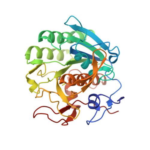
A detailed analysis of structural and intensity changes induced by X-ray radiation is presented for two types of proteinase K crystals: crystal grown by classical hanging drop method and those grown by Langmuir-Blodgett (LB) nanotemplate. The comparison of various parameters (e.g. intensity per sigma ratio, unit-cell volume, number of unique reflections, B-factors) and electron density maps as a function of radiation dose, demonstrates that crystals, grown by the LB nanotemplate method, appear to be more resistant against radiation damage than crystals grown by the classical hanging drop method.
- Nanoworld Institute, CIRSDNNOB University of Genova, Genoa, Italy.
Organizational Affiliation: