3DAD | pdb_00003dad

Crystal structure of the N-terminal regulatory domains of the formin FHOD1


Experimental Data Snapshot

  • Method: X-RAY DIFFRACTION
  • Resolution: 2.30 Å
  • R-Value Free: 
    0.259 (Depositor), 0.259 (DCC) 
  • R-Value Work: 
    0.219 (Depositor), 0.218 (DCC) 

wwPDB Validation 3D Report Full Report

Validation slider image for 3DAD

This is version 1.3 of the entry. See complete history

Literature

The Human Formin FHOD1 Contains a Bipartite Structure of FH3 and GTPase-Binding Domains Required for Activation.

Schulte, A.Stolp, B.Schonichen, A.Pylypenko, O.Rak, A.Fackler, O.T.Geyer, M.

(2008) Structure 16: 1313-1323

  • DOI: https://doi.org/10.1016/j.str.2008.06.008
  • Primary Citation Related Structures: 
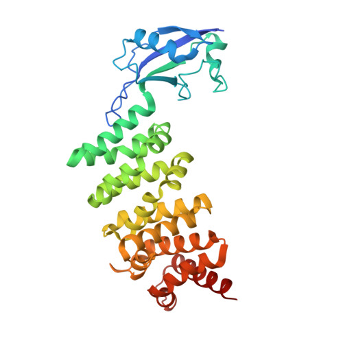
    3DAD

  • PubMed Abstract: 

    Formins induce the nucleation and polymerization of unbranched actin filaments. They share three homology domains required for profilin binding, actin polymerization, and regulation. Diaphanous-related formins (DRFs) are activated by GTPases of the Rho/Rac family, whose interaction with the N-terminal formin domain is thought to displace a C-terminal Diaphanous-autoregulatory domain (DAD). We have determined the structure of the N-terminal domains of FHOD1 consisting of a GTPase-binding domain (GBD) and the DAD-recognition domain FH3. In contrast to the formin mDia1, the FHOD1-GBD reveals a ubiquitin superfold as found similarly in c-Raf1 or PI3 kinase. This GBD is recruited by Rac and Ras GTPases in cells and plays an essential role for FHOD1-mediated actin remodeling. The FHOD1-FH3 domain is composed of five armadillo repeats, similarly to other formins. Mutation of one residue in the predicted DAD-interaction surface efficiently activates FHOD1 in cells. These results demonstrate that DRFs have evolved different molecular solutions to govern their autoregulation and GTPase specificity.


  • Organizational Affiliation
    • Abteilung Physikalische Biochemie, Max-Planck-Institut für Molekulare Physiologie, Otto-Hahn-Strasse 11, 44227 Dortmund, Germany.

Macromolecule Content 

  • Total Structure Weight: 74.55 kDa 
  • Atom Count: 5,072 
  • Modeled Residue Count: 635 
  • Deposited Residue Count: 678 
  • Unique protein chains: 1

Macromolecules

Find similar proteins by:|  3D Structure
Entity ID: 1
MoleculeChains  Sequence LengthOrganismDetailsImage
FH1/FH2 domain-containing protein 1
A, B
339Homo sapiensMutation(s): 2 
Gene Names: FHOD1FHOSFHOS1
UniProt & NIH Common Fund Data Resources
Find proteins for Q9Y613 (Homo sapiens)
Explore Q9Y613 
Go to UniProtKB:  Q9Y613
PHAROS:  Q9Y613
GTEx:  ENSG00000135723 
Entity Groups
Sequence Clusters30% Identity50% Identity70% Identity90% Identity95% Identity100% Identity
UniProt GroupQ9Y613
Sequence Annotations
Expand
Reference Sequence

Experimental Data & Validation

Experimental Data

  • Method: X-RAY DIFFRACTION
  • Resolution: 2.30 Å
  • R-Value Free:  0.259 (Depositor), 0.259 (DCC) 
  • R-Value Work:  0.219 (Depositor), 0.218 (DCC) 
Space Group: P 1
Unit Cell:
Length ( Å )Angle ( ˚ )
a = 35.444α = 78.24
b = 73.931β = 86.17
c = 78.674γ = 89.67
Software Package:
Software NamePurpose
CNSrefinement
CNSphasing

Structure Validation

View Full Validation Report


Entry History 

Deposition Data

Revision History  (Full details and data files)

  • Version 1.0: 2008-09-16
    Type: Initial release
  • Version 1.1: 2011-07-13
    Changes: Version format compliance
  • Version 1.2: 2021-10-20
    Changes: Database references
  • Version 1.3: 2024-02-21
    Changes: Data collection