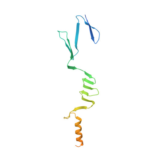
Structure of the head of the Bartonella adhesin BadA
Szczesny, P., Linke, D., Ursinus, A., Bar, K., Schwarz, H., Riess, T.M., Kempf, V.A., Lupas, A.N., Martin, J., Zeth, K.(2008) PLoS Pathog 4: e1000119-e1000119
- PubMed: 18688279 Search on PubMedSearch on PubMed Central
- DOI: https://doi.org/10.1371/journal.ppat.1000119
- Primary Citation Related Structures:
3D9X - PubMed Abstract:
Trimeric autotransporter adhesins (TAAs) are a major class of proteins by which pathogenic proteobacteria adhere to their hosts. Prominent examples include Yersinia YadA, Haemophilus Hia and Hsf, Moraxella UspA1 and A2, and Neisseria NadA. TAAs also occur in symbiotic and environmental species and presumably represent a general solution to the problem of adhesion in proteobacteria. The general structure of TAAs follows a head-stalk-anchor architecture, where the heads are the primary mediators of attachment and autoagglutination. In the major adhesin of Bartonella henselae, BadA, the head consists of three domains, the N-terminal of which shows strong sequence similarity to the head of Yersinia YadA. The two other domains were not recognizably similar to any protein of known structure. We therefore determined their crystal structure to a resolution of 1.1 A. Both domains are beta-prisms, the N-terminal one formed by interleaved, five-stranded beta-meanders parallel to the trimer axis and the C-terminal one by five-stranded beta-meanders orthogonal to the axis. Despite the absence of statistically significant sequence similarity, the two domains are structurally similar to domains from Haemophilus Hia, albeit in permuted order. Thus, the BadA head appears to be a chimera of domains seen in two other TAAs, YadA and Hia, highlighting the combinatorial evolutionary strategy taken by pathogens.
- Department of Protein Evolution, Max Planck Institute for Developmental Biology, Tübingen, Germany.
Organizational Affiliation: