3D72

1.65 Angstrom crystal structure of the Cys71Val variant in the fungal photoreceptor VVD


Experimental Data Snapshot

  • Method: X-RAY DIFFRACTION
  • Resolution: 1.65 Å
  • R-Value Free: 0.260 
  • R-Value Work: 0.250 
  • R-Value Observed: 0.260 

Starting Model: experimental
View more details

wwPDB Validation   3D Report Full Report


Ligand Structure Quality Assessment 


This is version 1.3 of the entry. See complete history


Literature

Light activation of the LOV protein vivid generates a rapidly exchanging dimer.

Zoltowski, B.D.Crane, B.R.

(2008) Biochemistry 47: 7012-7019

  • DOI: https://doi.org/10.1021/bi8007017
  • Primary Citation of Related Structures:  
    3D72

  • PubMed Abstract: 

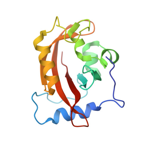
    The fungal photoreceptor Vivid (VVD) plays an important role in the adaptation of blue-light responses in Neurospora crassa. VVD, an FAD-binding LOV (light, oxygen, voltage) protein, couples light-induced cysteinyl adduct formation at the flavin ring to conformational changes in the N-terminal cap (Ncap) of the VVD PAS domain. Size-exclusion chromatography (SEC), equilibrium ultracentrifugation, and static and dynamic light scattering show that these conformational changes generate a rapidly exchanging VVD dimer, with an expanded hydrodynamic radius. A three-residue N-terminal beta-turn that assumes two different conformations in a crystal structure of a VVD C71V variant is essential for light-state dimerization. Residue substitutions at a critical hinge between the Ncap and PAS core can inhibit or enhance dimerization, whereas a Tyr to Trp substitution at the Ncap-PAS interface stabilizes the light-state dimer. Cross-linking through engineered disulfides indicates that the light-state dimer differs considerably from the dark-state dimer found in VVD crystal structures. These results verify the role of Ncap conformational changes in gating the photic response of N. crassa and indicate that LOV-LOV homo- or heterodimerization may be a mechanism for regulating light-activated gene expression.


  • Organizational Affiliation

    Department of Chemistry and Chemical Biology, Cornell University, Ithaca, New York 14853, USA.


Macromolecules
Find similar proteins by:  (by identity cutoff)  |  3D Structure
Entity ID: 1
MoleculeChains Sequence LengthOrganismDetailsImage
Vivid PAS protein VVD
A, B
149Neurospora crassaMutation(s): 1 
Gene Names: vvdG17A4.050
UniProt
Find proteins for Q1K5Y8 (Neurospora crassa (strain ATCC 24698 / 74-OR23-1A / CBS 708.71 / DSM 1257 / FGSC 987))
Explore Q1K5Y8 
Go to UniProtKB:  Q1K5Y8
Entity Groups  
Sequence Clusters30% Identity50% Identity70% Identity90% Identity95% Identity100% Identity
UniProt GroupQ1K5Y8
Sequence Annotations
Expand
  • Reference Sequence
Small Molecules
Ligands 1 Unique
IDChains Name / Formula / InChI Key2D Diagram3D Interactions
FAD
Query on FAD

Download Ideal Coordinates CCD File 
C [auth A],
D [auth B]
FLAVIN-ADENINE DINUCLEOTIDE
C27 H33 N9 O15 P2
VWWQXMAJTJZDQX-UYBVJOGSSA-N
Experimental Data & Validation

Experimental Data

  • Method: X-RAY DIFFRACTION
  • Resolution: 1.65 Å
  • R-Value Free: 0.260 
  • R-Value Work: 0.250 
  • R-Value Observed: 0.260 
  • Space Group: P 1 21 1
Unit Cell:
Length ( Å )Angle ( ˚ )
a = 36.24α = 90
b = 80.7β = 95.16
c = 57.58γ = 90
Software Package:
Software NamePurpose
HKL-2000data collection
AMoREphasing
CNSrefinement
HKL-2000data reduction
HKL-2000data scaling

Structure Validation

View Full Validation Report


Ligand Structure Quality Assessment 


Entry History 

Deposition Data

Revision History  (Full details and data files)

  • Version 1.0: 2008-06-17
    Type: Initial release
  • Version 1.1: 2011-07-13
    Changes: Version format compliance
  • Version 1.2: 2018-06-27
    Changes: Data collection, Database references
  • Version 1.3: 2023-08-30
    Changes: Data collection, Database references, Derived calculations, Refinement description