3D57

TR Variant D355R


Experimental Data Snapshot

  • Method: X-RAY DIFFRACTION
  • Resolution: 2.20 Å
  • R-Value Free: 0.240 
  • R-Value Work: 0.210 
  • R-Value Observed: 0.210 

wwPDB Validation   3D Report Full Report


Ligand Structure Quality Assessment 


This is version 1.3 of the entry. See complete history


Literature

Molecular basis for dimer formation of TRbeta variant D355R.

Jouravel, N.Sablin, E.Togashi, M.Baxter, J.D.Webb, P.Fletterick, R.J.

(2008) Proteins 75: 111-117

  • DOI: https://doi.org/10.1002/prot.22225
  • Primary Citation of Related Structures:  
    3D57

  • PubMed Abstract: 

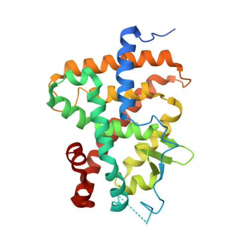
    Protein quality and stability are critical during protein purification for X-ray crystallography. A target protein that is easy to manipulate and crystallize becomes a valuable product useful for high-throughput crystallography for drug design and discovery. In this work, a single surface mutation, D355R, was shown to be crucial for converting the modestly stable monomeric ligand binding domain of the human thyroid hormone receptor (TR LBD) into a stable dimer. The structure of D335R TR LBD mutant was solved using X-ray crystallography and refined to 2.2 A resolution with R(free)/R values of 24.5/21.7. The crystal asymmetric unit reveals the TR dimer with two molecules of the hormone-bound LBD related by twofold symmetry. The ionic interface between the two LBDs comprises residues within loop H10-H11 and loop H6-H7 as well as the C-terminal halves of helices 8 of both protomers. Direct intermolecular contacts formed between the introduced residue Arg 355 of one TR molecule and Glu 324 of the second molecule become a part of the extended dimerization interface of 1330 A(2) characteristic for a strong complex assembly that is additionally strengthened by buffer solutes.


  • Organizational Affiliation

    Department of Biochemistry and Biophysics, University of California San Francisco (UCSF), San Francisco, California 94158, USA. njouravel@msg.ucsf.edu


Macromolecules
Find similar proteins by:  (by identity cutoff)  |  3D Structure
Entity ID: 1
MoleculeChains Sequence LengthOrganismDetailsImage
Thyroid hormone receptor beta
A, B
266Homo sapiensMutation(s): 1 
Gene Names: 7068ERBA2NR1A2THR1THRB
UniProt & NIH Common Fund Data Resources
Find proteins for P10828 (Homo sapiens)
Explore P10828 
Go to UniProtKB:  P10828
PHAROS:  P10828
GTEx:  ENSG00000151090 
Entity Groups  
Sequence Clusters30% Identity50% Identity70% Identity90% Identity95% Identity100% Identity
UniProt GroupP10828
Sequence Annotations
Expand
  • Reference Sequence
Small Molecules
Binding Affinity Annotations 
IDSourceBinding Affinity
4HY BindingDB:  3D57 IC50: min: 0.04, max: 0.15 (nM) from 4 assay(s)
Experimental Data & Validation

Experimental Data

  • Method: X-RAY DIFFRACTION
  • Resolution: 2.20 Å
  • R-Value Free: 0.240 
  • R-Value Work: 0.210 
  • R-Value Observed: 0.210 
  • Space Group: P 1 21 1
Unit Cell:
Length ( Å )Angle ( ˚ )
a = 55.557α = 90
b = 92.963β = 110.02
c = 58.808γ = 90
Software Package:
Software NamePurpose
ADSCdata collection
CNSrefinement
DENZOdata reduction
SCALEPACKdata scaling
CNSphasing

Structure Validation

View Full Validation Report


Ligand Structure Quality Assessment 


Entry History 

Deposition Data

  • Released Date: 2008-10-14 
  • Deposition Author(s): Jouravel, N.

Revision History  (Full details and data files)

  • Version 1.0: 2008-10-14
    Type: Initial release
  • Version 1.1: 2011-07-13
    Changes: Version format compliance
  • Version 1.2: 2021-10-20
    Changes: Database references, Derived calculations
  • Version 1.3: 2023-08-30
    Changes: Data collection, Refinement description