3D32 | pdb_00003d32

Complex of GABA(A) receptor-associated protein (GABARAP) with a synthetic peptide


Experimental Data Snapshot

  • Method: X-RAY DIFFRACTION
  • Resolution: 1.30 Å
  • R-Value Free: 
    0.195 (Depositor), 0.199 (DCC) 
  • R-Value Work: 
    0.177 (Depositor), 0.181 (DCC) 
  • R-Value Observed: 
    0.178 (Depositor) 

Starting Model: experimental
View more details

wwPDB Validation 3D Report Full Report

Validation slider image for 3D32

This is version 2.2 of the entry. See complete history

Literature

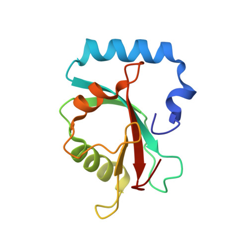
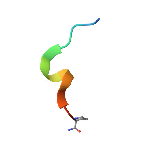
Ligand Binding Mode of GABA(A) Receptor-Associated Protein.

Weiergraber, O.H.Stangler, T.Thielmann, Y.Mohrluder, J.Wiesehan, K.Willbold, D.

(2008) J Mol Biology 381: 1320-1331

  • DOI: https://doi.org/10.1016/j.jmb.2008.06.086
  • Primary Citation Related Structures: 
    3D32

  • PubMed Abstract: 

    The gamma-aminobutyric acid type A (GABA(A)) receptor-associated protein is a versatile adaptor protein playing an important role in intracellular vesicle trafficking, particularly in neuronal cells. We present the X-ray structure of the soluble form of human GABA(A) receptor-associated protein complexed with a high-affinity synthetic peptide at 1.3 A resolution. The data shed light on the probable binding modes of key interaction partners, including the GABA(A) receptor and the cysteine protease Atg4. The resulting models provide a structural background for further investigation of the unique biological properties of this protein.


  • Organizational Affiliation
    • Institut für Neurowissenschaften und Biophysik, Molekulare Biophysik, Forschungszentrum Jülich, D-52425 Jülich, Germany.

Macromolecule Content 

  • Total Structure Weight: 31.46 kDa 
  • Atom Count: 2,599 
  • Modeled Residue Count: 260 
  • Deposited Residue Count: 264 
  • Unique protein chains: 2

Macromolecules

Find similar proteins by:|  3D Structure
Entity ID: 1
MoleculeChains  Sequence LengthOrganismDetailsImage
Gamma-aminobutyric acid receptor-associated protein
A, B
119Homo sapiensMutation(s): 0 
Gene Names: GABARAPFLC3BHT004
UniProt & NIH Common Fund Data Resources
Find proteins for O95166 (Homo sapiens)
Explore O95166 
Go to UniProtKB:  O95166
PHAROS:  O95166
GTEx:  ENSG00000170296 
Entity Groups
Sequence Clusters30% Identity50% Identity70% Identity90% Identity95% Identity100% Identity
UniProt GroupO95166
Sequence Annotations
Expand
Reference Sequence
Find similar proteins by:  Sequence   |   3D Structure  
Entity ID: 2
MoleculeChains  Sequence LengthOrganismDetailsImage
K1 peptide
C, D
13synthetic constructMutation(s): 0 

Small Molecules

Ligands 2 Unique
IDChains Name / Formula / InChI Key2D Diagram3D Interactions
CL

Query on CL


Download:Ideal Coordinates CCD File
F [auth A]
G [auth A]
H [auth A]
I [auth A]
J [auth B]
F [auth A],
G [auth A],
H [auth A],
I [auth A],
J [auth B],
K [auth B],
L [auth B],
M [auth B]
CHLORIDE ION
Cl
VEXZGXHMUGYJMC-UHFFFAOYSA-M
NA

Query on NA


Download:Ideal Coordinates CCD File
E [auth A]SODIUM ION
Na
FKNQFGJONOIPTF-UHFFFAOYSA-N

Experimental Data & Validation

Experimental Data

  • Method: X-RAY DIFFRACTION
  • Resolution: 1.30 Å
  • R-Value Free:  0.195 (Depositor), 0.199 (DCC) 
  • R-Value Work:  0.177 (Depositor), 0.181 (DCC) 
  • R-Value Observed: 0.178 (Depositor) 
Space Group: C 1 2 1
Unit Cell:
Length ( Å )Angle ( ˚ )
a = 129.287α = 90
b = 35.415β = 100.59
c = 58.209γ = 90
Software Package:
Software NamePurpose
MOSFLMdata reduction
SCALAdata scaling
MOLREPphasing
REFMACrefinement
PDB_EXTRACTdata extraction

Structure Validation

View Full Validation Report


Entry History 

Deposition Data

Revision History  (Full details and data files)

  • Version 1.0: 2008-08-05
    Type: Initial release
  • Version 1.1: 2011-07-13
    Changes: Advisory, Version format compliance
  • Version 2.0: 2020-03-04
    Changes: Atomic model, Data collection, Database references, Derived calculations, Polymer sequence, Source and taxonomy, Structure summary
  • Version 2.1: 2023-08-30
    Changes: Data collection, Database references, Derived calculations, Refinement description
  • Version 2.2: 2024-11-13
    Changes: Structure summary