3D17 | pdb_00003d17

A triply ligated crystal structure of relaxed state human hemoglobin


Experimental Data Snapshot

  • Method: X-RAY DIFFRACTION
  • Resolution: 2.80 Å
  • R-Value Free: 
    0.297 (Depositor), 0.299 (DCC) 
  • R-Value Work: 
    0.232 (Depositor), 0.229 (DCC) 
  • R-Value Observed: 
    0.255 (Depositor) 

Starting Model: experimental
View more details

wwPDB Validation 3D Report Full Report

Validation slider image for 3D17

Ligand Structure Quality Assessment 


This is version 1.3 of the entry. See complete history

Literature

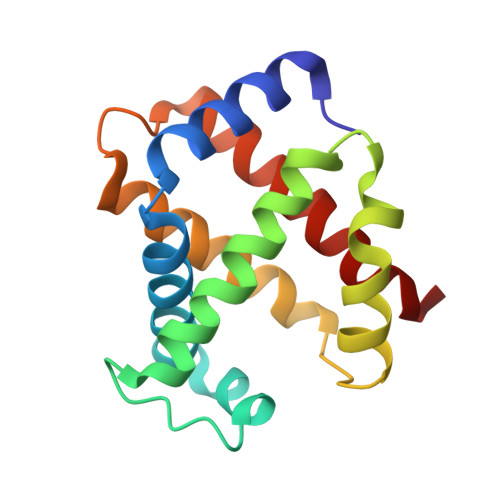
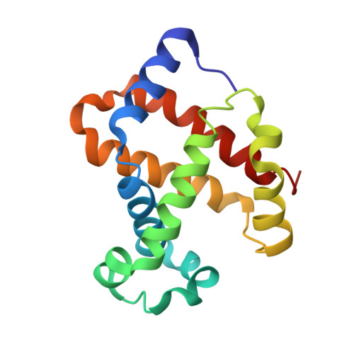
Structure of relaxed-state human hemoglobin: insight into ligand uptake, transport and release.

Jenkins, J.D.Musayev, F.N.Danso-Danquah, R.Abraham, D.J.Safo, M.K.

(2009) Acta Crystallogr D Biol Crystallogr 65: 41-48

  • DOI: https://doi.org/10.1107/S0907444908037256
  • Primary Citation Related Structures: 
    3D17

  • PubMed Abstract: 

    Hemoglobin was one of the first protein structures to be determined by X-ray crystallography and served as a basis for the two-state MWC model for the mechanism of allosteric proteins. Since then, there has been an ongoing debate about whether Hb allostery involves the unliganded tense T state and the liganded relaxed R state or whether it involves the T state and an ensemble of liganded relaxed states. In fact, the former model is inconsistent with many functional observations, as well as the recent discoveries of several relaxed-state Hb structures such as RR2, R3 and R2. One school of thought has suggested the R2 state to be the physiologically relevant relaxed end state, with the R state mediating the T-->R2 transition. X-ray studies have been performed on human carbonmonoxy Hb at a resolution of 2.8 A. The ensuing liganded quaternary structure is different from previously reported liganded Hb structures. The distal beta-heme pocket is the largest when compared with other liganded Hb structures, partly owing to rotation of betaHis63(E7) out of the distal pocket, creating a ligand channel to the solvent. The structure also shows unusually smaller alpha- and beta-clefts. Results from this study taken in conjunction with previous findings suggest that multiple liganded Hb states with different quaternary structures may be involved in ligand uptake, stabilization, transport and release.


  • Organizational Affiliation
    • Department of Medicinal Chemistry, School of Pharmacy and Institute for Structural Biology and Drug Discovery, Virginia Commonwealth University, Richmond, VA 23219, USA.

Macromolecule Content 

  • Total Structure Weight: 65.01 kDa 
  • Atom Count: 4,672 
  • Modeled Residue Count: 574 
  • Deposited Residue Count: 574 
  • Unique protein chains: 2

Macromolecules

Find similar proteins by:|  3D Structure
Entity ID: 1
MoleculeChains  Sequence LengthOrganismDetailsImage
Hemoglobin subunit alpha
A, C
141Homo sapiensMutation(s): 0 
UniProt & NIH Common Fund Data Resources
Find proteins for P69905 (Homo sapiens)
Explore P69905 
Go to UniProtKB:  P69905
Entity Groups
Sequence Clusters30% Identity50% Identity70% Identity90% Identity95% Identity100% Identity
UniProt GroupP69905
Sequence Annotations
Expand
Reference Sequence
Find similar proteins by:|  3D Structure
Entity ID: 2
MoleculeChains  Sequence LengthOrganismDetailsImage
Hemoglobin subunit beta
B, D
146Homo sapiensMutation(s): 0 
UniProt & NIH Common Fund Data Resources
Find proteins for P68871 (Homo sapiens)
Explore P68871 
Go to UniProtKB:  P68871
PHAROS:  P68871
GTEx:  ENSG00000244734 
Entity Groups
Sequence Clusters30% Identity50% Identity70% Identity90% Identity95% Identity100% Identity
UniProt GroupP68871
Sequence Annotations
Expand
Reference Sequence

Small Molecules

Ligands 4 Unique
IDChains Name / Formula / InChI Key2D Diagram3D Interactions
HEM

Query on HEM


Download:Ideal Coordinates CCD File
E [auth A],
J [auth B],
L [auth C],
N [auth D]
PROTOPORPHYRIN IX CONTAINING FE
C34 H32 Fe N4 O4
KABFMIBPWCXCRK-RGGAHWMASA-L
PO4

Query on PO4


Download:Ideal Coordinates CCD File
H [auth B],
I [auth B],
M [auth D]
PHOSPHATE ION
O4 P
NBIIXXVUZAFLBC-UHFFFAOYSA-K
MBN

Query on MBN


Download:Ideal Coordinates CCD File
F [auth A]TOLUENE
C7 H8
YXFVVABEGXRONW-UHFFFAOYSA-N
CMO

Query on CMO


Download:Ideal Coordinates CCD File
G [auth A],
K [auth B],
O [auth D]
CARBON MONOXIDE
C O
UGFAIRIUMAVXCW-UHFFFAOYSA-N

Experimental Data & Validation

Experimental Data

  • Method: X-RAY DIFFRACTION
  • Resolution: 2.80 Å
  • R-Value Free:  0.297 (Depositor), 0.299 (DCC) 
  • R-Value Work:  0.232 (Depositor), 0.229 (DCC) 
  • R-Value Observed: 0.255 (Depositor) 
Space Group: P 41 21 2
Unit Cell:
Length ( Å )Angle ( ˚ )
a = 87.282α = 90
b = 87.282β = 90
c = 174.466γ = 90
Software Package:
Software NamePurpose
d*TREKdata scaling
AMoREphasing
CNSrefinement
PDB_EXTRACTdata extraction
d*TREKdata reduction

Structure Validation

View Full Validation Report


Ligand Structure Quality Assessment 


Entry History 

Deposition Data

Revision History  (Full details and data files)

  • Version 1.0: 2008-06-03
    Type: Initial release
  • Version 1.1: 2011-07-13
    Changes: Version format compliance
  • Version 1.2: 2023-08-30
    Changes: Data collection, Database references, Derived calculations, Refinement description
  • Version 1.3: 2025-11-12
    Changes: Database references, Structure summary