3D0Y

Crystal Structure of S100B in the Calcium and Zinc Loaded State at pH 6.5


Experimental Data Snapshot

  • Method: X-RAY DIFFRACTION
  • Resolution: 1.50 Å
  • R-Value Free: 0.261 
  • R-Value Work: 0.206 
  • R-Value Observed: 0.208 

wwPDB Validation   3D Report Full Report


This is version 1.2 of the entry. See complete history


Literature

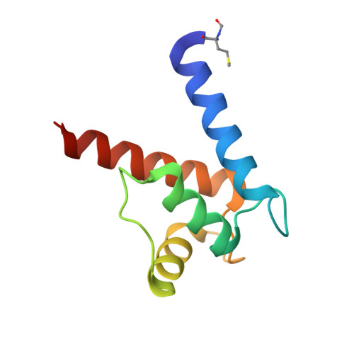
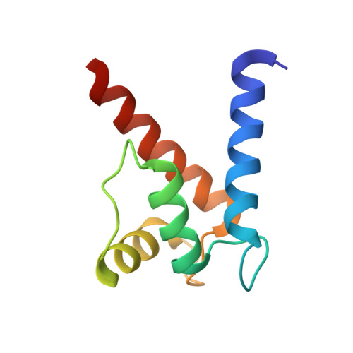
The crystal structures of human S100B in the zinc- and calcium-loaded state at three pH values reveal zinc ligand swapping.

Ostendorp, T.Diez, J.Heizmann, C.W.Fritz, G.

(2011) Biochim Biophys Acta 1813: 1083-1091

  • DOI: https://doi.org/10.1016/j.bbamcr.2010.10.006
  • Primary Citation of Related Structures:  
    3CZT, 3D0Y, 3D10

  • PubMed Abstract: 

    S100B is a homodimeric zinc-, copper-, and calcium-binding protein of the family of EF-hand S100 proteins. Zn(2+) binding to S100B increases its affinity towards Ca(2+) as well as towards target peptides and proteins. Cu(2+) and Zn(2+) bind presumably to the same site in S100B. We determined the structures of human Zn(2+)- and Ca(2+)-loaded S100B at pH 6.5, pH 9, and pH 10 by X-ray crystallography at 1.5, 1.4, and 1.65Å resolution, respectively. Two Zn(2+) ions are coordinated tetrahedrally at the dimer interface by His and Glu residues from both subunits. The crystal structures revealed that ligand swapping occurs for one of the four ligands in the Zn(2+)-binding sites. Whereas at pH 9, the Zn(2+) ions are coordinated by His15, His25, His 85', and His 90', at pH 6.5 and pH 10, His90' is replaced by Glu89'. The results document that the Zn(2+)-binding sites are flexible to accommodate other metal ions such as Cu(2+). Moreover, we characterized the structural changes upon Zn(2+) binding, which might lead to increased affinity towards Ca(2+) as well as towards target proteins. We observed that in Zn(2+)-Ca(2+)-loaded S100B the C-termini of helix IV adopt a distinct conformation. Zn(2+) binding induces a repositioning of residues Phe87 and Phe88, which are involved in target protein binding. This article is part of a Special Issue entitled: 11th European Symposium on Calcium.


  • Organizational Affiliation

    Fachbereich Biologie, Mathematisch-Naturwissenschaftliche Sektion, Universität Konstanz, 78459 Konstanz, Germany.


Macromolecules
Find similar proteins by:  (by identity cutoff)  |  3D Structure
Entity ID: 1
MoleculeChains Sequence LengthOrganismDetailsImage
Protein S100-B92Homo sapiensMutation(s): 0 
Gene Names: S100B
UniProt & NIH Common Fund Data Resources
Find proteins for P04271 (Homo sapiens)
Explore P04271 
Go to UniProtKB:  P04271
PHAROS:  P04271
GTEx:  ENSG00000160307 
Entity Groups  
Sequence Clusters30% Identity50% Identity70% Identity90% Identity95% Identity100% Identity
UniProt GroupP04271
Sequence Annotations
Expand
  • Reference Sequence
Find similar proteins by:  (by identity cutoff)  |  3D Structure
Entity ID: 2
MoleculeChains Sequence LengthOrganismDetailsImage
Protein S100-B92Homo sapiensMutation(s): 0 
Gene Names: S100B
UniProt & NIH Common Fund Data Resources
Find proteins for P04271 (Homo sapiens)
Explore P04271 
Go to UniProtKB:  P04271
PHAROS:  P04271
GTEx:  ENSG00000160307 
Entity Groups  
Sequence Clusters30% Identity50% Identity70% Identity90% Identity95% Identity100% Identity
UniProt GroupP04271
Sequence Annotations
Expand
  • Reference Sequence
Experimental Data & Validation

Experimental Data

  • Method: X-RAY DIFFRACTION
  • Resolution: 1.50 Å
  • R-Value Free: 0.261 
  • R-Value Work: 0.206 
  • R-Value Observed: 0.208 
  • Space Group: P 1 21 1
Unit Cell:
Length ( Å )Angle ( ˚ )
a = 34.89α = 90
b = 58.15β = 111.12
c = 47.66γ = 90
Software Package:
Software NamePurpose
XSCALEdata scaling
MOLREPphasing
REFMACrefinement
PDB_EXTRACTdata extraction
XDSdata reduction

Structure Validation

View Full Validation Report


Entry History 

Deposition Data

Revision History  (Full details and data files)

  • Version 1.0: 2009-04-14
    Type: Initial release
  • Version 1.1: 2011-07-13
    Changes: Advisory, Version format compliance
  • Version 1.2: 2023-08-30
    Changes: Data collection, Database references, Derived calculations, Refinement description