3D0E | pdb_00003d0e

Crystal structure of human Akt2 in complex with GSK690693


Experimental Data Snapshot

  • Method: X-RAY DIFFRACTION
  • Resolution: 2.00 Å
  • R-Value Free: 
    0.246 (Depositor), 0.246 (DCC) 
  • R-Value Work: 
    0.206 (Depositor), 0.209 (DCC) 
  • R-Value Observed: 
    0.208 (Depositor) 

wwPDB Validation 3D Report Full Report

Validation slider image for 3D0E

Ligand Structure Quality Assessment 


This is version 1.4 of the entry. See complete history

Literature

Identification of 4-(2-(4-amino-1,2,5-oxadiazol-3-yl)-1-ethyl-7-{[(3S)-3-piperidinylmethyl]oxy}-1H-imidazo[4,5-c]pyridin-4-yl)-2-methyl-3-butyn-2-ol (GSK690693), a novel inhibitor of AKT kinase.

Heerding, D.A.Rhodes, N.Leber, J.D.Clark, T.J.Keenan, R.M.Lafrance, L.V.Li, M.Safonov, I.G.Takata, D.T.Venslavsky, J.W.Yamashita, D.S.Choudhry, A.E.Copeland, R.A.Lai, Z.Schaber, M.D.Tummino, P.J.Strum, S.L.Wood, E.R.Duckett, D.R.Eberwein, D.Knick, V.B.Lansing, T.J.McConnell, R.T.Zhang, S.Minthorn, E.A.Concha, N.O.Warren, G.L.Kumar, R.

(2008) J Med Chem 51: 5663-5679

  • DOI: https://doi.org/10.1021/jm8004527
  • Primary Citation Related Structures: 
    3D0E

  • PubMed Abstract: 

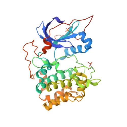
    Overexpression of AKT has an antiapoptotic effect in many cell types, and expression of dominant negative AKT blocks the ability of a variety of growth factors to promote survival. Therefore, inhibitors of AKT kinase activity might be useful as monotherapy for the treatment of tumors with activated AKT. Herein, we describe our lead optimization studies culminating in the discovery of compound 3g (GSK690693). Compound 3g is a novel ATP competitive, pan-AKT kinase inhibitor with IC 50 values of 2, 13, and 9 nM against AKT1, 2, and 3, respectively. An X-ray cocrystal structure was solved with 3g and the kinase domain of AKT2, confirming that 3g bound in the ATP binding pocket. Compound 3g potently inhibits intracellular AKT activity as measured by the inhibition of the phosphorylation levels of GSK3beta. Intraperitoneal administration of 3g in immunocompromised mice results in the inhibition of GSK3beta phosphorylation and tumor growth in human breast carcinoma (BT474) xenografts.


  • Organizational Affiliation
    • Oncology Center of Excellence for Drug Discovery, GlaxoSmithKline, 1250 South Collegeville Road, Collegeville, Pennsylvania 19426, USA. dirk.a.heerding@gsk.com

Macromolecule Content 

  • Total Structure Weight: 79 kDa 
  • Atom Count: 5,826 
  • Modeled Residue Count: 648 
  • Deposited Residue Count: 670 
  • Unique protein chains: 1

Macromolecules

Find similar proteins by:|  3D Structure
Entity ID: 1
MoleculeChains  Sequence LengthOrganismDetailsImage
RAC-beta serine/threonine-protein kinase
A, B
335Homo sapiensMutation(s): 1 
Gene Names: AKT2
EC: 2.7.11.1
UniProt & NIH Common Fund Data Resources
Find proteins for P31751 (Homo sapiens)
Explore P31751 
Go to UniProtKB:  P31751
PHAROS:  P31751
GTEx:  ENSG00000105221 
Entity Groups
Sequence Clusters30% Identity50% Identity70% Identity90% Identity95% Identity100% Identity
UniProt GroupP31751
Sequence Annotations
Expand
Reference Sequence

Small Molecules

Ligands 1 Unique
IDChains Name / Formula / InChI Key2D Diagram3D Interactions
G93

Query on G93


Download:Ideal Coordinates CCD File
C [auth A],
D [auth B]
4-{2-(4-amino-1,2,5-oxadiazol-3-yl)-1-ethyl-7-[(3S)-piperidin-3-ylmethoxy]-1H-imidazo[4,5-c]pyridin-4-yl}-2-methylbut-3 -yn-2-ol
C21 H27 N7 O3
KGPGFQWBCSZGEL-ZDUSSCGKSA-N
Modified Residues  1 Unique
IDChains TypeFormula2D DiagramParent
TPO
Query on TPO
A, B
L-PEPTIDE LINKINGC4 H10 N O6 PTHR

Experimental Data & Validation

Experimental Data

  • Method: X-RAY DIFFRACTION
  • Resolution: 2.00 Å
  • R-Value Free:  0.246 (Depositor), 0.246 (DCC) 
  • R-Value Work:  0.206 (Depositor), 0.209 (DCC) 
  • R-Value Observed: 0.208 (Depositor) 
Space Group: P 32
Unit Cell:
Length ( Å )Angle ( ˚ )
a = 116.489α = 90
b = 116.489β = 90
c = 45.119γ = 120
Software Package:
Software NamePurpose
DENZOdata reduction
SCALEPACKdata scaling
REFMACrefinement
PDB_EXTRACTdata extraction

Structure Validation

View Full Validation Report


Ligand Structure Quality Assessment 


Entry History 

Deposition Data

Revision History  (Full details and data files)

  • Version 1.0: 2008-10-21
    Type: Initial release
  • Version 1.1: 2011-07-13
    Changes: Advisory, Version format compliance
  • Version 1.2: 2016-12-28
    Changes: Non-polymer description
  • Version 1.3: 2017-10-25
    Changes: Refinement description
  • Version 1.4: 2024-10-16
    Changes: Data collection, Database references, Derived calculations, Refinement description, Structure summary