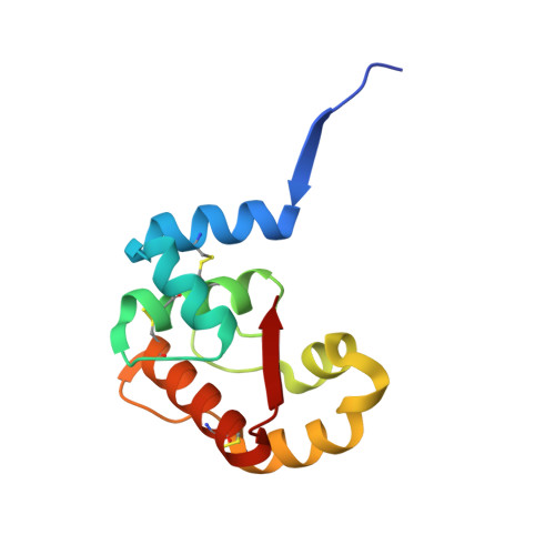
3CYZ

Dimeric crystal structure of a pheromone binding protein from Apis mellifera in complex with 9-keto-2(E)-decenoic acid at pH 7.0


Experimental Data Snapshot

  • Method: X-RAY DIFFRACTION
  • Resolution: 1.80 Å
  • R-Value Free: 0.200 
  • R-Value Work: 0.162 
  • R-Value Observed: 0.164 

Starting Model: experimental
View more details

wwPDB Validation   3D Report Full Report


Ligand Structure Quality Assessment 


This is version 1.2 of the entry. See complete history


Literature

Queen bee pheromone binding protein pH-induced domain swapping favors pheromone release

Pesenti, M.E.Spinelli, S.Bezirard, V.Briand, L.Pernollet, J.C.Campanacci, V.Tegoni, M.Cambillau, C.

(2009) J Mol Biol 390: 981-990

  • DOI: https://doi.org/10.1016/j.jmb.2009.05.067
  • Primary Citation of Related Structures:  
    3CYZ, 3CZ0, 3CZ1, 3CZ2, 3D73, 3D74, 3D75, 3D76, 3D77, 3D78

  • PubMed Abstract: 

    In honeybee (Apis mellifera) societies, the queen controls the development and the caste status of the members of the hive. Queen bees secrete pheromonal blends comprising 10 or more major and minor components, mainly hydrophobic. The major component, 9-keto-2(E)-decenoic acid (9-ODA), acts on the workers and male bees (drones), eliciting social or sexual responses. 9-ODA is captured in the antennal lymph and transported to the pheromone receptor(s) in the sensory neuron membranes by pheromone binding proteins (PBPs). A key issue is to understand how the pheromone, once tightly bound to its PBP, is released to activate the receptor. We report here on the structure at physiological pH of the main antennal PBP, ASP1, identified in workers and male honeybees, in its apo or complexed form, particularly with the main component of the queen mandibular pheromonal mixture (9-ODA). Contrary to the ASP1 structure at low pH, the ASP1 structure at pH 7.0 is a domain-swapped dimer with one or two ligands per monomer. This dimerization is disrupted by a unique residue mutation since Asp35 Asn and Asp35 Ala mutants remain monomeric at pH 7.0, as does native ASP1 at pH 4.0. Asp35 is conserved in only approximately 30% of medium-chain PBPs and is replaced by other residues, such as Asn, Ala and Ser, among others, thus excluding that they may perform domain swapping. Therefore, these different medium-chain PBPs, as well as PBPs from moths, very likely exhibit different mechanisms of ligand release or receptor recognition.


  • Organizational Affiliation

    Architecture et Fonction des Macromolécules Biologiques, UMR CNRS, Marseille, France.


Macromolecules
Find similar proteins by:  (by identity cutoff)  |  3D Structure
Entity ID: 1
MoleculeChains Sequence LengthOrganismDetailsImage
Pheromone-binding protein ASP1
A, B
119Apis melliferaMutation(s): 0 
UniProt
Find proteins for Q9U9J6 (Apis mellifera)
Explore Q9U9J6 
Go to UniProtKB:  Q9U9J6
Entity Groups  
Sequence Clusters30% Identity50% Identity70% Identity90% Identity95% Identity100% Identity
UniProt GroupQ9U9J6
Sequence Annotations
Expand
  • Reference Sequence
Small Molecules
Ligands 4 Unique
IDChains Name / Formula / InChI Key2D Diagram3D Interactions
9OD
Query on 9OD

Download Ideal Coordinates CCD File 
C [auth A],
H [auth B],
I [auth B]
(2Z)-9-oxodec-2-enoic acid
C10 H16 O3
INJRDZMWIAYEMM-VURMDHGXSA-N
GOL
Query on GOL

Download Ideal Coordinates CCD File 
K [auth B],
L [auth B]
GLYCEROL
C3 H8 O3
PEDCQBHIVMGVHV-UHFFFAOYSA-N
CL
Query on CL

Download Ideal Coordinates CCD File 
D [auth A],
E [auth A],
F [auth A],
J [auth B]
CHLORIDE ION
Cl
VEXZGXHMUGYJMC-UHFFFAOYSA-M
MG
Query on MG

Download Ideal Coordinates CCD File 
G [auth A]MAGNESIUM ION
Mg
JLVVSXFLKOJNIY-UHFFFAOYSA-N
Binding Affinity Annotations 
IDSourceBinding Affinity
9OD PDBBind:  3CYZ Kd: 60 (nM) from 1 assay(s)
Experimental Data & Validation

Experimental Data

  • Method: X-RAY DIFFRACTION
  • Resolution: 1.80 Å
  • R-Value Free: 0.200 
  • R-Value Work: 0.162 
  • R-Value Observed: 0.164 
  • Space Group: P 21 21 21
Unit Cell:
Length ( Å )Angle ( ˚ )
a = 36.285α = 90
b = 75.555β = 90
c = 84.113γ = 90
Software Package:
Software NamePurpose
REFMACrefinement
ADSCdata collection
MOSFLMdata reduction
SCALAdata scaling
AMoREphasing

Structure Validation

View Full Validation Report


Ligand Structure Quality Assessment 


Entry History 

Deposition Data

Revision History  (Full details and data files)

  • Version 1.0: 2009-04-28
    Type: Initial release
  • Version 1.1: 2011-07-13
    Changes: Advisory, Version format compliance
  • Version 1.2: 2023-11-01
    Changes: Data collection, Database references, Derived calculations, Refinement description