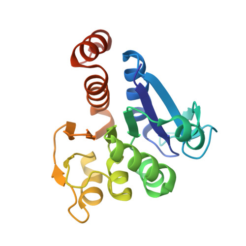
Cysteine pKa depression by a protonated glutamic acid in human DJ-1.
Witt, A.C., Lakshminarasimhan, M., Remington, B.C., Hasim, S., Pozharski, E., Wilson, M.A.(2008) Biochemistry 47: 7430-7440
- PubMed: 18570440
- DOI: https://doi.org/10.1021/bi800282d
- Primary Citation of Related Structures:
2OR3, 3CY6, 3CYF, 3CZ9, 3CZA - PubMed Abstract:
Human DJ-1, a disease-associated protein that protects cells from oxidative stress, contains an oxidation-sensitive cysteine (C106) that is essential for its cytoprotective activity. The origin of C106 reactivity is obscure, due in part to the absence of an experimentally determined p K a value for this residue. We have used atomic-resolution X-ray crystallography and UV spectroscopy to show that C106 has a depressed p K a of 5.4 +/- 0.1 and that the C106 thiolate accepts a hydrogen bond from a protonated glutamic acid side chain (E18). X-ray crystal structures and cysteine p K a analysis of several site-directed substitutions at residue 18 demonstrate that the protonated carboxylic acid side chain of E18 is required for the maximal stabilization of the C106 thiolate. A nearby arginine residue (R48) participates in a guanidinium stacking interaction with R28 from the other monomer in the DJ-1 dimer and elevates the p K a of C106 by binding an anion that electrostatically suppresses thiol ionization. Our results show that the ionizable residues (E18, R48, and R28) surrounding C106 affect its p K a in a way that is contrary to expectations based on the typical ionization behavior of glutamic acid and arginine. Lastly, a search of the Protein Data Bank (PDB) produces several candidate hydrogen-bonded aspartic/glutamic acid-cysteine interactions, which we propose are particularly common in the DJ-1 superfamily.
- Department of Biochemistry and Redox Biology Center, The University of Nebraska, Lincoln, Nebraska 68588-0664, USA.
Organizational Affiliation:


