3CVZ | pdb_00003cvz

Structural insights into the molecular organization of the S-layer from Clostridium difficile


Experimental Data Snapshot

  • Method: X-RAY DIFFRACTION
  • Resolution: 2.40 Å
  • R-Value Free: 
    0.294 (Depositor), 0.287 (DCC) 
  • R-Value Work: 
    0.231 (Depositor), 0.227 (DCC) 
  • R-Value Observed: 
    0.233 (Depositor) 

wwPDB Validation 3D Report Full Report

Validation slider image for 3CVZ

This is version 1.3 of the entry. See complete history

Literature

Structural insights into the molecular organization of the S-layer from Clostridium difficile

Fagan, R.P.Albesa-Jove, D.Qazi, O.Svergun, D.I.Brown, K.A.Fairweather, N.F.

(2009) Mol Microbiol 71: 1308-1322

  • DOI: https://doi.org/10.1111/j.1365-2958.2009.06603.x
  • Primary Citation Related Structures: 
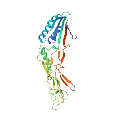
    3CVZ

  • PubMed Abstract: 

    Clostridium difficile expresses a surface layer (S-layer) which coats the surface of the bacterium and acts as an adhesin facilitating interaction of the bacterium with host enteric cells. The S-layer contains a high-molecular-weight S-layer protein (HMW SLP) and its low-molecular-weight partner protein (LMW SLP). We show that these proteins form a tightly associated non-covalent complex, the H/L complex, and we identify the regions of both proteins responsible for complex formation. The 2.4 A X-ray crystal structure of a truncated derivative of the LMW SLP reveals two domains. Domain 1 has a two-layer sandwich architecture while domain 2, predicted to orientate towards the external environment, contains a novel fold. Small-angle X-ray scattering analysis of the H/L complex shows an elongated molecule, with the two SLPs arranged 'end-to-end' interacting with each other through a small contact area. Alignment of LMW SLPs, which exhibit high sequence diversity, reveals a core of conserved residues that could reflect functional conservation, while allowing for immune evasion through sequence variation. These structures are the first described for the S-layer of a bacterial pathogen, and provide insights into the assembly and biogenesis of the S-layer.


  • Organizational Affiliation
    • Division of Cell and Molecular Biology, Centre for Molecular Microbiology and Infection, Imperial College London, London SW72AZ, UK.

Macromolecule Content 

  • Total Structure Weight: 116.49 kDa 
  • Atom Count: 7,641 
  • Modeled Residue Count: 976 
  • Deposited Residue Count: 1,092 
  • Unique protein chains: 1

Macromolecules

Find similar proteins by:|  3D Structure
Entity ID: 1
MoleculeChains  Sequence LengthOrganismDetailsImage
S-layer protein
A, B, C, D
273Clostridioides difficileMutation(s): 0 
Gene Names: slpA
UniProt
Find proteins for Q183M8 (Clostridioides difficile (strain 630))
Explore Q183M8 
Go to UniProtKB:  Q183M8
Entity Groups
Sequence Clusters30% Identity50% Identity70% Identity90% Identity95% Identity100% Identity
UniProt GroupQ183M8
Sequence Annotations
Expand
Reference Sequence

Experimental Data & Validation

Experimental Data

  • Method: X-RAY DIFFRACTION
  • Resolution: 2.40 Å
  • R-Value Free:  0.294 (Depositor), 0.287 (DCC) 
  • R-Value Work:  0.231 (Depositor), 0.227 (DCC) 
  • R-Value Observed: 0.233 (Depositor) 
Space Group: P 64
Unit Cell:
Length ( Å )Angle ( ˚ )
a = 107.059α = 90
b = 107.059β = 90
c = 190.617γ = 120
Software Package:
Software NamePurpose
DENZOdata reduction
SCALEPACKdata scaling
SHARPphasing
PHENIXrefinement
PDB_EXTRACTdata extraction
MAR345data collection

Structure Validation

View Full Validation Report


Entry History 

Deposition Data

Revision History  (Full details and data files)

  • Version 1.0: 2009-03-17
    Type: Initial release
  • Version 1.1: 2011-07-13
    Changes: Version format compliance
  • Version 1.2: 2017-10-25
    Changes: Refinement description
  • Version 1.3: 2024-02-21
    Changes: Data collection, Database references, Derived calculations