3CVM

High resolution structure of a stable Plasminogen activator inhibitor type-1 in its protease cleaved form


Experimental Data Snapshot

  • Method: X-RAY DIFFRACTION
  • Resolution: 2.02 Å
  • R-Value Free: 0.282 
  • R-Value Work: 0.228 
  • R-Value Observed: 0.229 

wwPDB Validation   3D Report Full Report


This is version 1.3 of the entry. See complete history


Literature

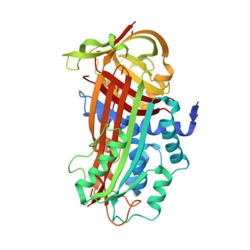
High-resolution structure of the stable plasminogen activator inhibitor type-1 variant 14-1B in its proteinase-cleaved form: a new tool for detailed interaction studies and modeling.

Jensen, J.K.Gettins, P.G.

(2008) Protein Sci 17: 1844-1849

  • DOI: https://doi.org/10.1110/ps.036707.108
  • Primary Citation of Related Structures:  
    3CVM

  • PubMed Abstract: 

    Wild-type plasminogen activator inhibitor type-1 (PAI-1) rapidly converts to the inactive latent state under conditions of physiological pH and temperature. For in vivo studies of active PAI-1 in cell culture and in vivo model systems, the 14-1B PAI-1 mutant (N150H-K154T-Q319L-M354I), with its stabilized active conformation, has thus become the PAI-1 of choice. As a consequence of the increased stability, the only two forms likely to be encountered are the active or the cleaved form, the latter either free or complexed with target proteinase. We hereby report the first structure of the stable 14-1B PAI-1 variant in its reactive center cleaved form, to a resolution of 2.0 A. The >99% complete structure represents the highest resolved structure of free cleaved PAI-1. This high-resolution structure should be of great use for drug target development and for modeling protein-protein interactions such as those of PAI-1 with vitronectin.


  • Organizational Affiliation

    Department of Biochemistry and Molecular Genetics, University of Illinois at Chicago, Chicago, Illinois 60607, USA.


Macromolecules
Find similar proteins by:  (by identity cutoff)  |  3D Structure
Entity ID: 1
MoleculeChains Sequence LengthOrganismDetailsImage
Plasminogen activator inhibitor 1
A, B
392Homo sapiensMutation(s): 5 
Gene Names: SERPINE1PAI1PLANH1
UniProt & NIH Common Fund Data Resources
Find proteins for P05121 (Homo sapiens)
Explore P05121 
Go to UniProtKB:  P05121
PHAROS:  P05121
GTEx:  ENSG00000106366 
Entity Groups  
Sequence Clusters30% Identity50% Identity70% Identity90% Identity95% Identity100% Identity
UniProt GroupP05121
Sequence Annotations
Expand
  • Reference Sequence
Experimental Data & Validation

Experimental Data

  • Method: X-RAY DIFFRACTION
  • Resolution: 2.02 Å
  • R-Value Free: 0.282 
  • R-Value Work: 0.228 
  • R-Value Observed: 0.229 
  • Space Group: P 1 21 1
Unit Cell:
Length ( Å )Angle ( ˚ )
a = 76.603α = 90
b = 104.014β = 101.93
c = 76.426γ = 90
Software Package:
Software NamePurpose
XSCALEdata processing
PHENIXrefinement
PDB_EXTRACTdata extraction
MAR345dtbdata collection
XDSdata reduction
XSCALEdata scaling
PHASERphasing

Structure Validation

View Full Validation Report


Entry History 

Deposition Data

Revision History  (Full details and data files)

  • Version 1.0: 2008-08-19
    Type: Initial release
  • Version 1.1: 2011-07-13
    Changes: Version format compliance
  • Version 1.2: 2021-10-20
    Changes: Database references
  • Version 1.3: 2023-08-30
    Changes: Data collection, Refinement description