


Truncated APS kinase from Pencillium chrysogenum: Insight into the function of the N-terminal helix
Gay, S.C., Segel, I.H., Fisher, A.J.To be published.
Experimental Data Snapshot
Starting Model: experimental
View more details
Entity ID: 1 | |||||
|---|---|---|---|---|---|
| Molecule | Chains | Sequence Length | Organism | Details | Image |
| Adenylyl-sulfate kinase | 197 | Penicillium chrysogenum | Mutation(s): 0 EC: 2.7.1.25 | 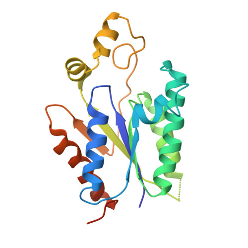 | |
UniProt | |||||
Entity Groups | |||||
| Sequence Clusters | 30% Identity50% Identity70% Identity90% Identity95% Identity100% Identity | ||||
| UniProt Group | Q12657 | ||||
Sequence AnnotationsExpand | |||||
Reference Sequence | |||||
| Ligands 3 Unique | |||||
|---|---|---|---|---|---|
| ID | Chains | Name / Formula / InChI Key | 2D Diagram | 3D Interactions | |
| PPS Download:Ideal Coordinates CCD File | G [auth A], L [auth C] | 3'-PHOSPHATE-ADENOSINE-5'-PHOSPHATE SULFATE C10 H15 N5 O13 P2 S GACDQMDRPRGCTN-KQYNXXCUSA-N |  | ||
| ADP Download:Ideal Coordinates CCD File | F [auth A], I [auth B], K [auth C], M [auth D] | ADENOSINE-5'-DIPHOSPHATE C10 H15 N5 O10 P2 XTWYTFMLZFPYCI-KQYNXXCUSA-N |  | ||
| CL Download:Ideal Coordinates CCD File | E [auth A], H [auth B], J [auth C] | CHLORIDE ION Cl VEXZGXHMUGYJMC-UHFFFAOYSA-M |  | ||
| Length ( Å ) | Angle ( ˚ ) |
|---|---|
| a = 77.58 | α = 90 |
| b = 82.594 | β = 90 |
| c = 139.009 | γ = 90 |
| Software Name | Purpose |
|---|---|
| SCALA | data scaling |
| EPMR | phasing |
| PHENIX | refinement |
| PDB_EXTRACT | data extraction |
| Blu-Ice | data collection |
| MOSFLM | data reduction |


