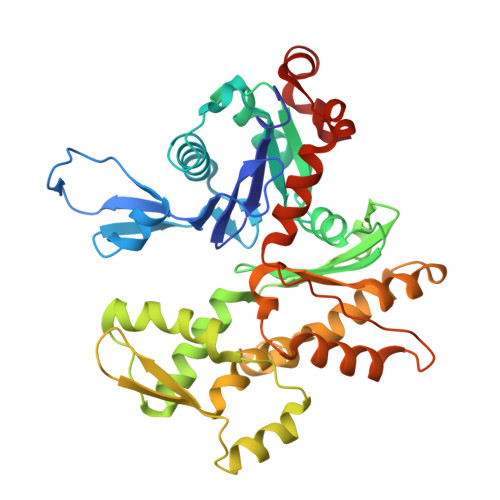
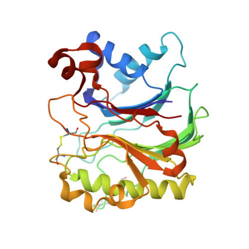
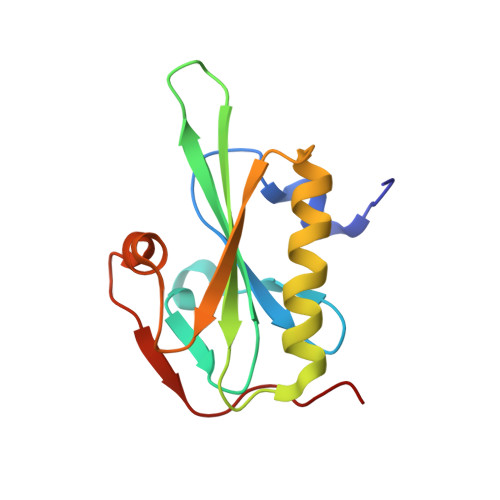
3CJC

Actin dimer cross-linked by V. cholerae MARTX toxin and complexed with DNase I and Gelsolin-segment 1


Experimental Data Snapshot

  • Method: X-RAY DIFFRACTION
  • Resolution: 3.90 Å
  • R-Value Free: 0.278 
  • R-Value Work: 0.223 
  • R-Value Observed: 0.226 

wwPDB Validation   3D Report Full Report


Ligand Structure Quality Assessment 


This is version 2.0 of the entry. See complete history


Literature

Connecting actin monomers by iso-peptide bond is a toxicity mechanism of the Vibrio cholerae MARTX toxin.

Kudryashov, D.S.Durer, Z.A.Ytterberg, A.J.Sawaya, M.R.Pashkov, I.Prochazkova, K.Yeates, T.O.Loo, R.R.Loo, J.A.Satchell, K.J.Reisler, E.

(2008) Proc Natl Acad Sci U S A 105: 18537-18542

  • DOI: https://doi.org/10.1073/pnas.0808082105
  • Primary Citation of Related Structures:  
    3CJB, 3CJC

  • PubMed Abstract: 

    The Gram-negative bacterium Vibrio cholerae is the causative agent of a severe diarrheal disease that afflicts three to five million persons annually, causing up to 200,000 deaths. Nearly all V. cholerae strains produce a large multifunctional-autoprocessing RTX toxin (MARTX(Vc)), which contributes significantly to the pathogenesis of cholera in model systems. The actin cross-linking domain (ACD) of MARTX(Vc) directly catalyzes a covalent cross-linking of monomeric G-actin into oligomeric chains and causes cell rounding, but the nature of the cross-linked bond and the mechanism of the actin cytoskeleton disruption remained elusive. To elucidate the mechanism of ACD action and effect on actin, we identified the covalent cross-link bond between actin protomers using limited proteolysis, X-ray crystallography, and mass spectrometry. We report here that ACD catalyzes the formation of an intermolecular iso-peptide bond between residues E270 and K50 located in the hydrophobic and the DNaseI-binding loops of actin, respectively. Mutagenesis studies confirm that no other residues on actin can be cross-linked by ACD both in vitro and in vivo. This cross-linking locks actin protomers into an orientation different from that of F-actin, resulting in strong inhibition of actin polymerization. This report describes a microbial toxin mechanism acting via iso-peptide bond cross-linking between host proteins and is, to the best of our knowledge, the only known example of a peptide linkage between nonterminal glutamate and lysine side chains.


  • Organizational Affiliation

    Department of Chemistry and Biochemistry and Molecular Biology Institute, University of California, Los Angeles, CA 90095, USA.


Macromolecules
Find similar proteins by:  (by identity cutoff)  |  3D Structure
Entity ID: 1
MoleculeChains Sequence LengthOrganismDetailsImage
Actin, alpha skeletal muscle377Oryctolagus cuniculusMutation(s): 0 
UniProt
Find proteins for P68135 (Oryctolagus cuniculus)
Explore P68135 
Go to UniProtKB:  P68135
Entity Groups  
Sequence Clusters30% Identity50% Identity70% Identity90% Identity95% Identity100% Identity
UniProt GroupP68135
Sequence Annotations
Expand
  • Reference Sequence
Find similar proteins by:  (by identity cutoff)  |  3D Structure
Entity ID: 2
MoleculeChains Sequence LengthOrganismDetailsImage
Deoxyribonuclease-1B [auth D]260Bos taurusMutation(s): 0 
EC: 3.1.21.1
UniProt
Find proteins for P00639 (Bos taurus)
Explore P00639 
Go to UniProtKB:  P00639
Entity Groups  
Sequence Clusters30% Identity50% Identity70% Identity90% Identity95% Identity100% Identity
UniProt GroupP00639
Sequence Annotations
Expand
  • Reference Sequence
Find similar proteins by:  (by identity cutoff)  |  3D Structure
Entity ID: 3
MoleculeChains Sequence LengthOrganismDetailsImage
GelsolinC [auth G]125Homo sapiensMutation(s): 0 
Gene Names: GSN
UniProt & NIH Common Fund Data Resources
Find proteins for P06396 (Homo sapiens)
Explore P06396 
Go to UniProtKB:  P06396
PHAROS:  P06396
GTEx:  ENSG00000148180 
Entity Groups  
Sequence Clusters30% Identity50% Identity70% Identity90% Identity95% Identity100% Identity
UniProt GroupP06396
Sequence Annotations
Expand
  • Reference Sequence
Oligosaccharides

Help

Entity ID: 4
MoleculeChains Length2D Diagram Glycosylation3D Interactions
2-acetamido-2-deoxy-beta-D-glucopyranose-(1-4)-2-acetamido-2-deoxy-beta-D-glucopyranoseD [auth B]2N-Glycosylation
Glycosylation Resources
GlyTouCan:  G42666HT
GlyCosmos:  G42666HT
GlyGen:  G42666HT
Small Molecules
Ligands 3 Unique
IDChains Name / Formula / InChI Key2D Diagram3D Interactions
ATP
Query on ATP

Download Ideal Coordinates CCD File 
G [auth A]ADENOSINE-5'-TRIPHOSPHATE
C10 H16 N5 O13 P3
ZKHQWZAMYRWXGA-KQYNXXCUSA-N
SO4
Query on SO4

Download Ideal Coordinates CCD File 
F [auth A]
H [auth D]
I [auth D]
J [auth D]
K [auth D]
F [auth A],
H [auth D],
I [auth D],
J [auth D],
K [auth D],
L [auth G]
SULFATE ION
O4 S
QAOWNCQODCNURD-UHFFFAOYSA-L
CA
Query on CA

Download Ideal Coordinates CCD File 
E [auth A],
M [auth G]
CALCIUM ION
Ca
BHPQYMZQTOCNFJ-UHFFFAOYSA-N
Experimental Data & Validation

Experimental Data

  • Method: X-RAY DIFFRACTION
  • Resolution: 3.90 Å
  • R-Value Free: 0.278 
  • R-Value Work: 0.223 
  • R-Value Observed: 0.226 
  • Space Group: P 21 21 21
Unit Cell:
Length ( Å )Angle ( ˚ )
a = 89.303α = 90
b = 108.566β = 90
c = 133.761γ = 90
Software Package:
Software NamePurpose
DENZOdata reduction
SCALEPACKdata scaling
PHASERphasing
REFMACrefinement
PDB_EXTRACTdata extraction
BOSdata collection

Structure Validation

View Full Validation Report


Ligand Structure Quality Assessment 


Entry History 

Deposition Data

Revision History  (Full details and data files)

  • Version 1.0: 2008-03-25
    Type: Initial release
  • Version 1.1: 2011-07-13
    Changes: Advisory, Version format compliance
  • Version 1.2: 2017-10-25
    Changes: Refinement description
  • Version 2.0: 2020-07-29
    Type: Remediation
    Reason: Carbohydrate remediation
    Changes: Atomic model, Data collection, Derived calculations, Structure summary