


Site-Selective Glycosylation of Cysteine-93 beta on the Surface of Bovine Hemoglobin and its Application as a Novel Oxygen Therapeutic
Bhatt, V.S., Zhang, Y., Sun, G., Wang, P.G., Palmer, A.F.To be published.
Experimental Data Snapshot
Starting Model: experimental
View more details
Entity ID: 1 | |||||
|---|---|---|---|---|---|
| Molecule | Chains | Sequence Length | Organism | Details | Image |
| Hemoglobin subunit alpha | 141 | Bos taurus | Mutation(s): 0 Gene Names: HBA | 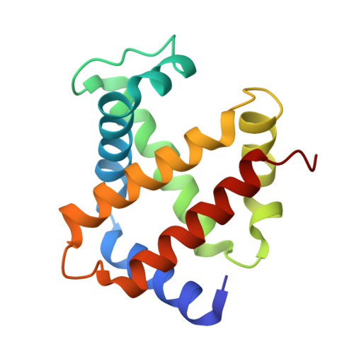 | |
UniProt | |||||
Entity Groups | |||||
| Sequence Clusters | 30% Identity50% Identity70% Identity90% Identity95% Identity100% Identity | ||||
| UniProt Group | P01966 | ||||
Sequence AnnotationsExpand | |||||
Reference Sequence | |||||
Entity ID: 2 | |||||
|---|---|---|---|---|---|
| Molecule | Chains | Sequence Length | Organism | Details | Image |
| Hemoglobin subunit beta | 145 | Bos taurus | Mutation(s): 0 Gene Names: HBB | 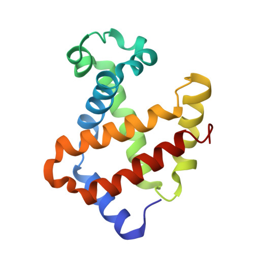 | |
UniProt | |||||
Entity Groups | |||||
| Sequence Clusters | 30% Identity50% Identity70% Identity90% Identity95% Identity100% Identity | ||||
| UniProt Group | P02070 | ||||
Sequence AnnotationsExpand | |||||
Reference Sequence | |||||
| Ligands 3 Unique | |||||
|---|---|---|---|---|---|
| ID | Chains | Name / Formula / InChI Key | 2D Diagram | 3D Interactions | |
| 5DP Download:Ideal Coordinates CCD File | I [auth B] | 5-(2,5-dioxopyrrolidin-1-yl)-N-[2-(2-{2-[(4-O-alpha-D-idopyranosyl-alpha-D-mannopyranosyl)oxy]ethoxy}ethoxy)ethyl]pentanamide C27 H46 N2 O16 GIIWJKKPIBKANP-SLHLUNFVSA-N |  | ||
| HEM Download:Ideal Coordinates CCD File | E [auth A], G [auth B], J [auth C], L [auth D] | PROTOPORPHYRIN IX CONTAINING FE C34 H32 Fe N4 O4 KABFMIBPWCXCRK-RGGAHWMASA-L |  | ||
| O Download:Ideal Coordinates CCD File | F [auth A], H [auth B], K [auth C], M [auth D] | OXYGEN ATOM O XLYOFNOQVPJJNP-UHFFFAOYSA-N |  | ||
| Length ( Å ) | Angle ( ˚ ) |
|---|---|
| a = 62.604 | α = 90 |
| b = 73.982 | β = 90 |
| c = 129.796 | γ = 90 |
| Software Name | Purpose |
|---|---|
| REFMAC | refinement |
| CrystalClear | data collection |
| CrystalClear | data reduction |
| CrystalClear | data scaling |
| AMoRE | phasing |


