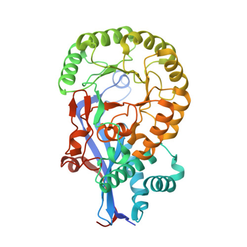
Crystal structure of L-Talarate dehydratase from Polaromonas sp. JS666 complexed with Mg and L-glucarate.
Fedorov, A.A., Fedorov, E.V., Yew, W.S., Burley, S.K., Gerlt, J.A., Almo, S.C.To be published.
Experimental Data Snapshot
| Ligands 2 Unique | |||||
|---|---|---|---|---|---|
| ID | Chains | Name / Formula / InChI Key | 2D Diagram | 3D Interactions | |
| LGT Download:Ideal Coordinates CCD File | G [auth A], J [auth B], M [auth C], P [auth D] | L-GLUCARIC ACID C6 H10 O8 DSLZVSRJTYRBFB-AJSXGEPRSA-N |  | ||
| MG Download:Ideal Coordinates CCD File | E [auth A] F [auth A] H [auth B] I [auth B] K [auth C] | MAGNESIUM ION Mg JLVVSXFLKOJNIY-UHFFFAOYSA-N |  | ||
| Length ( Å ) | Angle ( ˚ ) |
|---|---|
| a = 195.466 | α = 90 |
| b = 84.739 | β = 126.04 |
| c = 118.247 | γ = 90 |
| Software Name | Purpose |
|---|---|
| CNS | refinement |
| ADSC | data collection |
| DENZO | data reduction |
| SCALEPACK | data scaling |
| SOLVE | phasing |