3C9E

Crystal structure of the cathepsin K : chondroitin sulfate complex.


Experimental Data Snapshot

  • Method: X-RAY DIFFRACTION
  • Resolution: 1.80 Å
  • R-Value Free: 0.209 
  • R-Value Work: 0.180 
  • R-Value Observed: 0.180 

wwPDB Validation   3D Report Full Report


Ligand Structure Quality Assessment 


This is version 2.0 of the entry. See complete history


Literature

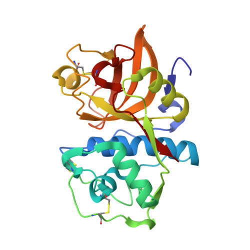
The crystal and molecular structures of a cathepsin K:chondroitin sulfate complex.

Li, Z.Kienetz, M.Cherney, M.M.James, M.N.Bromme, D.

(2008) J Mol Biol 383: 78-91

  • DOI: https://doi.org/10.1016/j.jmb.2008.07.038
  • Primary Citation of Related Structures:  
    3C9E

  • PubMed Abstract: 

    Cathepsin K is the major collagenolytic enzyme produced by bone-resorbing osteoclasts. We showed earlier that the unique triple-helical collagen-degrading activity of cathepsin K depends on the formation of complexes with bone-or cartilage-resident glycosaminoglycans, such as chondroitin 4-sulfate (C4-S). Here, we describe the crystal structure of a 1:n complex of cathepsin K:C4-S inhibited by E64 at a resolution of 1.8 A. The overall structure reveals an unusual "beads-on-a-string"-like organization. Multiple cathepsin K molecules bind specifically to a single cosine curve-shaped strand of C4-S with each cathepsin K molecule interacting with three disaccharide residues of C4-S. One of the more important sets of interactions comes from a single turn of helix close to the N terminus of the proteinase containing a basic amino acid triplet (Arg8-Lys9-Lys10) that forms multiple hydrogen bonds either to the caboxylate or to the 4-sulfate groups of C4-S. Altogether, the binding sites with C4-S are located in the R-domain of cathepsin K and are distant from its active site. This explains why the general proteolytic activity of cathepsin K is not affected by the binding of chondroitin sulfate. Biochemical analyses of cathepsin K and C4-S mixtures support the presence of a 1:n complex in solution; a dissociation constant, K(d), of about 10 nM was determined for the interaction between cathepsin K and C4-S.


  • Organizational Affiliation

    Mount Sinai School of Medicine, Department of Human Genetics, New York, NY 10029, USA.


Macromolecules
Find similar proteins by:  (by identity cutoff)  |  3D Structure
Entity ID: 1
MoleculeChains Sequence LengthOrganismDetailsImage
Cathepsin K215Homo sapiensMutation(s): 0 
Gene Names: CTSKCTSOCTSO2
EC: 3.4.22.38
UniProt & NIH Common Fund Data Resources
Find proteins for P43235 (Homo sapiens)
Explore P43235 
Go to UniProtKB:  P43235
PHAROS:  P43235
GTEx:  ENSG00000143387 
Entity Groups  
Sequence Clusters30% Identity50% Identity70% Identity90% Identity95% Identity100% Identity
UniProt GroupP43235
Sequence Annotations
Expand
  • Reference Sequence
Oligosaccharides

Help

Entity ID: 2
MoleculeChains Length2D Diagram Glycosylation3D Interactions
2-acetamido-2-deoxy-4-O-sulfo-beta-D-galactopyranose-(1-4)-beta-D-glucopyranuronic acid-(1-3)-2-acetamido-2-deoxy-4-O-sulfo-beta-D-galactopyranose-(1-4)-beta-D-glucopyranuronic acid-(1-3)-2-acetamido-2-deoxy-4-O-sulfo-beta-D-galactopyranose-(1-4)-beta-D-glucopyranuronic acid
B
6N/A
Glycosylation Resources
GlyTouCan:  G16831RV
GlyCosmos:  G16831RV
Small Molecules
Ligands 2 Unique
IDChains Name / Formula / InChI Key2D Diagram3D Interactions
E64
Query on E64

Download Ideal Coordinates CCD File 
D [auth A]N-[N-[1-HYDROXYCARBOXYETHYL-CARBONYL]LEUCYLAMINO-BUTYL]-GUANIDINE
C15 H30 N5 O5
QPQNJAXBPHVASB-QWRGUYRKSA-O
CA
Query on CA

Download Ideal Coordinates CCD File 
C [auth A]CALCIUM ION
Ca
BHPQYMZQTOCNFJ-UHFFFAOYSA-N
Binding Affinity Annotations 
IDSourceBinding Affinity
E64 PDBBind:  3C9E Kd: 10 (nM) from 1 assay(s)
Experimental Data & Validation

Experimental Data

  • Method: X-RAY DIFFRACTION
  • Resolution: 1.80 Å
  • R-Value Free: 0.209 
  • R-Value Work: 0.180 
  • R-Value Observed: 0.180 
  • Space Group: C 2 2 21
Unit Cell:
Length ( Å )Angle ( ˚ )
a = 42α = 90
b = 143.9β = 90
c = 87.2γ = 90
Software Package:
Software NamePurpose
MOSFLMdata reduction
CNSrefinement
PDB_EXTRACTdata extraction
ADSCdata collection
SCALAdata scaling
MOLREPphasing

Structure Validation

View Full Validation Report


Ligand Structure Quality Assessment 


Entry History 

Deposition Data

Revision History  (Full details and data files)

  • Version 1.0: 2008-08-26
    Type: Initial release
  • Version 1.1: 2011-07-13
    Changes: Non-polymer description, Version format compliance
  • Version 1.2: 2016-02-17
    Changes: Derived calculations
  • Version 2.0: 2020-07-29
    Type: Remediation
    Reason: Carbohydrate remediation
    Changes: Atomic model, Data collection, Derived calculations, Structure summary