3C9A

High Resolution Crystal Structure of Argos bound to the EGF domain of Spitz


Experimental Data Snapshot

  • Method: X-RAY DIFFRACTION
  • Resolution: 1.60 Å
  • R-Value Free: 0.240 
  • R-Value Work: 0.195 
  • R-Value Observed: 0.198 

wwPDB Validation   3D Report Full Report


This is version 1.3 of the entry. See complete history


Literature

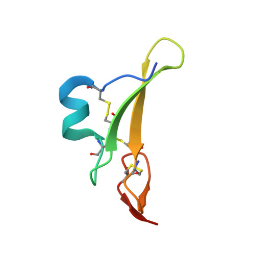
Structural basis for EGFR ligand sequestration by Argos.

Klein, D.E.Stayrook, S.E.Shi, F.Narayan, K.Lemmon, M.A.

(2008) Nature 453: 1271-1275

  • DOI: https://doi.org/10.1038/nature06978
  • Primary Citation of Related Structures:  
    3C9A, 3CA7, 3CGU

  • PubMed Abstract: 

    Members of the epidermal growth factor receptor (EGFR) or ErbB/HER family and their activating ligands are essential regulators of diverse developmental processes. Inappropriate activation of these receptors is a key feature of many human cancers, and its reversal is an important clinical goal. A natural secreted antagonist of EGFR signalling, called Argos, was identified in Drosophila. We showed previously that Argos functions by directly binding (and sequestering) growth factor ligands that activate EGFR. Here we describe the 1.6-A resolution crystal structure of Argos bound to an EGFR ligand. Contrary to expectations, Argos contains no EGF-like domain. Instead, a trio of closely related domains (resembling a three-finger toxin fold) form a clamp-like structure around the bound EGF ligand. Although structurally unrelated to the receptor, Argos mimics EGFR by using a bipartite binding surface to entrap EGF. The individual Argos domains share unexpected structural similarities with the extracellular ligand-binding regions of transforming growth factor-beta family receptors. The three-domain clamp of Argos also resembles the urokinase-type plasminogen activator (uPA) receptor, which uses a similar mechanism to engulf the EGF-like module of uPA. Our results indicate that undiscovered mammalian counterparts of Argos may exist among other poorly characterized structural homologues. In addition, the structures presented here define requirements for the design of artificial EGF-sequestering proteins that would be valuable anti-cancer therapeutics.


  • Organizational Affiliation

    Department of Biochemistry and Biophysics, University of Pennsylvania School of Medicine, 809C Stellar-Chance Laboratories, 422 Curie Boulevard, Philadelphia, Pennsylvania 19104-6059, USA.


Macromolecules
Find similar proteins by:  (by identity cutoff)  |  3D Structure
Entity ID: 1
MoleculeChains Sequence LengthOrganismDetailsImage
Protein giant-lens
A, B
223Drosophila melanogasterMutation(s): 0 
Gene Names: argosAosgilsty
UniProt
Find proteins for Q00805 (Drosophila melanogaster)
Explore Q00805 
Go to UniProtKB:  Q00805
Entity Groups  
Sequence Clusters30% Identity50% Identity70% Identity90% Identity95% Identity100% Identity
UniProt GroupQ00805
Sequence Annotations
Expand
  • Reference Sequence
Find similar proteins by:  (by identity cutoff)  |  3D Structure
Entity ID: 2
MoleculeChains Sequence LengthOrganismDetailsImage
Protein spitz
C, D
52Drosophila melanogasterMutation(s): 0 
Gene Names: spi
UniProt
Find proteins for Q01083 (Drosophila melanogaster)
Explore Q01083 
Go to UniProtKB:  Q01083
Entity Groups  
Sequence Clusters30% Identity50% Identity70% Identity90% Identity95% Identity100% Identity
UniProt GroupQ01083
Sequence Annotations
Expand
  • Reference Sequence
Small Molecules
Ligands 1 Unique
IDChains Name / Formula / InChI Key2D Diagram3D Interactions
BR
Query on BR

Download Ideal Coordinates CCD File 
E [auth A]
F [auth A]
G [auth A]
H [auth A]
I [auth A]
E [auth A],
F [auth A],
G [auth A],
H [auth A],
I [auth A],
J [auth A],
K [auth A],
L [auth A],
M [auth A],
N [auth B],
O [auth B],
P [auth B],
Q [auth B],
R [auth B],
S [auth B],
T [auth B],
U [auth C],
V [auth D]
BROMIDE ION
Br
CPELXLSAUQHCOX-UHFFFAOYSA-M
Experimental Data & Validation

Experimental Data

  • Method: X-RAY DIFFRACTION
  • Resolution: 1.60 Å
  • R-Value Free: 0.240 
  • R-Value Work: 0.195 
  • R-Value Observed: 0.198 
  • Space Group: P 1
Unit Cell:
Length ( Å )Angle ( ˚ )
a = 49.991α = 84.23
b = 51.275β = 74.82
c = 69.985γ = 75.67
Software Package:
Software NamePurpose
REFMACrefinement
HKL-2000data reduction
SCALEPACKdata scaling
SHELXCDphasing
SHELXEmodel building

Structure Validation

View Full Validation Report


Entry History 

Deposition Data

Revision History  (Full details and data files)

  • Version 1.0: 2008-05-20
    Type: Initial release
  • Version 1.1: 2011-07-13
    Changes: Version format compliance
  • Version 1.2: 2017-08-16
    Changes: Source and taxonomy
  • Version 1.3: 2017-10-25
    Changes: Refinement description