


Crystal structure of the periplasmic domain of Vibrio Cholerae LuxQ
Slama, B., Hendrickson, W.To be published.
Experimental Data Snapshot
wwPDB Validation 3D Report Full Report

Entity ID: 1 | |||||
|---|---|---|---|---|---|
| Molecule | Chains | Sequence Length | Organism | Details | Image |
| Autoinducer 2 sensor kinase/phosphatase luxQ | 270 | Vibrio cholerae | Mutation(s): 0 Gene Names: luxQ EC: 2.7.13.3 (PDB Primary Data), 3.1.3 (PDB Primary Data) | 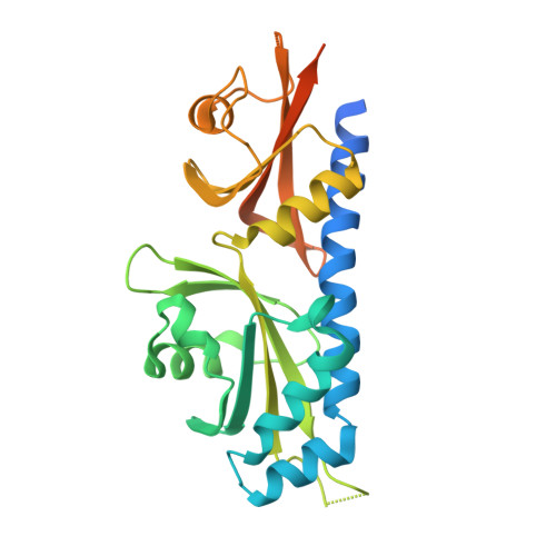 | |
UniProt | |||||
Entity Groups | |||||
| Sequence Clusters | 30% Identity50% Identity70% Identity90% Identity95% Identity100% Identity | ||||
| UniProt Group | Q9KLK7 | ||||
Sequence AnnotationsExpand | |||||
Reference Sequence | |||||
| Length ( Å ) | Angle ( ˚ ) |
|---|---|
| a = 55.656 | α = 90 |
| b = 55.656 | β = 90 |
| c = 161.415 | γ = 90 |
| Software Name | Purpose |
|---|---|
| CNS | refinement |
| HKL-2000 | data reduction |
| HKL-2000 | data scaling |
| SHARP | phasing |


