3C05 | pdb_00003c05

Crystal structure of Acostatin from Agkistrodon Contortrix Contortrix


Experimental Data Snapshot

  • Method: X-RAY DIFFRACTION
  • Resolution: 1.70 Å
  • R-Value Free: 
    0.215 (Depositor), 0.216 (DCC) 
  • R-Value Work: 
    0.186 (Depositor), 0.188 (DCC) 
  • R-Value Observed: 
    0.188 (Depositor) 

Starting Model: experimental
View more details

wwPDB Validation 3D Report Full Report

Validation slider image for 3C05

This is version 2.1 of the entry. See complete history

Literature

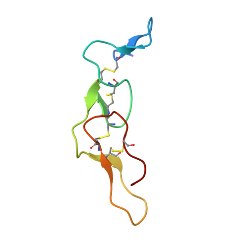
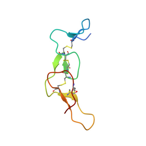
Crystal structure of acostatin, a dimeric disintegrin from southern copperhead (Agkistrodon contortrix contortrix) at 1.7 A resolution

Moiseeva, N.Bau, R.Swenson, S.D.Markland, F.S.Choe, J.-Y.Liu, Z.-J.Allaire, M.

(2008) Acta Crystallogr D Biol Crystallogr 64: 466-470

  • DOI: https://doi.org/10.1107/S0907444908002370
  • Primary Citation Related Structures: 
    3C05

  • PubMed Abstract: 

    Disintegrins are a family of small (4-14 kDa) proteins that bind to another class of proteins, integrins. Therefore, as integrin inhibitors, they can be exploited as anticancer and antiplatelet agents. Acostatin, an alphabeta heterodimeric disintegrin, has been isolated from the venom of Southern copperhead (Agkistrodon contortrix contortrix). The three-dimensional structure of acostatin has been determined by macromolecular crystallography using the molecular-replacement method. The asymmetric unit of the acostatin crystals consists of two heterodimers. The structure has been refined to an R(work) and R(free) of 18.6% and 21.5%, respectively, using all data in the 20-1.7 A resolution range. The structure of all subunits is similar and is well ordered into N-terminal and C-terminal clusters with four intramolecular disulfide bonds. The overall fold consists of short beta-sheets, each of which is formed by a pair of antiparallel beta-strands connected by beta-turns and flexible loops of different lengths. Conformational flexibility is found in the RGD loops and in the C-terminal segment. The interaction of two N-terminal clusters via two intermolecular disulfide bridges anchors the alphabeta chains of the acostatin dimers. The C-terminal clusters of the heterodimer project in opposite directions and form a larger angle between them in comparison with other dimeric disintegrins. Extensive interactions are observed between two heterodimers, revealing an alphabetabetaalpha acostatin tetramer. Further experiments are required to identify whether the alphabetabetaalpha acostatin complex plays a functional role in vivo.


  • Organizational Affiliation
    • National Synchrotron Light Source, Brookhaven National Laboratory, Building 725D, Upton, NY 11973, USA.

Macromolecule Content 

  • Total Structure Weight: 27.38 kDa 
  • Atom Count: 2,004 
  • Modeled Residue Count: 232 
  • Deposited Residue Count: 252 
  • Unique protein chains: 2

Macromolecules

Find similar proteins by:|  3D Structure
Entity ID: 1
MoleculeChains  Sequence LengthOrganismDetailsImage
Disintegrin acostatin alpha
A, C
62Agkistrodon contortrix contortrixMutation(s): 0 
UniProt
Find proteins for Q805F7 (Agkistrodon contortrix contortrix)
Explore Q805F7 
Go to UniProtKB:  Q805F7
Entity Groups
Sequence Clusters30% Identity50% Identity70% Identity90% Identity95% Identity100% Identity
UniProt GroupQ805F7
Sequence Annotations
Expand
Reference Sequence
Find similar proteins by:|  3D Structure
Entity ID: 2
MoleculeChains  Sequence LengthOrganismDetailsImage
Disintegrin acostatin-beta
B, D
64Agkistrodon contortrix contortrixMutation(s): 0 
UniProt
Find proteins for Q805F6 (Agkistrodon contortrix contortrix)
Explore Q805F6 
Go to UniProtKB:  Q805F6
Entity Groups
Sequence Clusters30% Identity50% Identity70% Identity90% Identity95% Identity100% Identity
UniProt GroupQ805F6
Sequence Annotations
Expand
Reference Sequence

Experimental Data & Validation

Experimental Data

  • Method: X-RAY DIFFRACTION
  • Resolution: 1.70 Å
  • R-Value Free:  0.215 (Depositor), 0.216 (DCC) 
  • R-Value Work:  0.186 (Depositor), 0.188 (DCC) 
  • R-Value Observed: 0.188 (Depositor) 
Space Group: P 21 21 21
Unit Cell:
Length ( Å )Angle ( ˚ )
a = 37.449α = 90
b = 59.808β = 90
c = 121.307γ = 90
Software Package:
Software NamePurpose
DENZOdata reduction
SCALEPACKdata scaling
REFMACrefinement
PDB_EXTRACTdata extraction
HKL-2000data reduction
HKL-2000data scaling
PHASERphasing

Structure Validation

View Full Validation Report


Entry History 

Deposition Data

Revision History  (Full details and data files)

  • Version 1.0: 2008-04-29
    Type: Initial release
  • Version 1.1: 2011-07-13
    Changes: Version format compliance
  • Version 1.2: 2017-10-25
    Changes: Refinement description
  • Version 2.0: 2023-08-30
    Changes: Data collection, Database references, Derived calculations, Polymer sequence, Refinement description
  • Version 2.1: 2024-10-30
    Changes: Structure summary