3BWU

Crystal structure of the ternary complex of FimD (N-Terminal Domain, FimDN) with FimC and the N-terminally truncated pilus subunit FimF (FimFt)


Experimental Data Snapshot

  • Method: X-RAY DIFFRACTION
  • Resolution: 1.76 Å
  • R-Value Free: 0.212 
  • R-Value Work: 0.175 

wwPDB Validation   3D Report Full Report


This is version 1.2 of the entry. See complete history


Literature

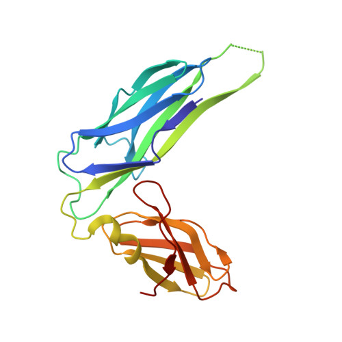
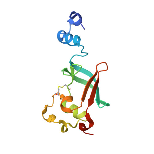
Crystal structure of the ternary FimC-FimF(t)-FimD(N) complex indicates conserved pilus chaperone-subunit complex recognition by the usher FimD

Eidam, O.Dworkowski, F.S.Glockshuber, R.Grutter, M.G.Capitani, G.

(2008) FEBS Lett 582: 651-655

  • DOI: https://doi.org/10.1016/j.febslet.2008.01.030
  • Primary Citation of Related Structures:  
    3BWU

  • PubMed Abstract: 
  • Type 1 pili, anchored to the outer membrane protein FimD, enable uropathogenic Escherichia coli to attach to host cells. During pilus biogenesis, the N-terminal periplasmic domain of FimD (FimD(N)) binds complexes between the chaperone FimC and pilus subunits via its partly disordered N-terminal segment, as recently shown for the FimC-FimH(P)-FimD(N) ternary complex ...

    Type 1 pili, anchored to the outer membrane protein FimD, enable uropathogenic Escherichia coli to attach to host cells. During pilus biogenesis, the N-terminal periplasmic domain of FimD (FimD(N)) binds complexes between the chaperone FimC and pilus subunits via its partly disordered N-terminal segment, as recently shown for the FimC-FimH(P)-FimD(N) ternary complex. We report the structure of a new ternary complex (FimC-FimF(t)-FimD(N)) with the subunit FimF(t) instead of FimH(p). FimD(N) recognizes FimC-FimF(t) and FimC-FimH(P) very similarly, predominantly through hydrophobic interactions. The conserved binding mode at a "hot spot" on the chaperone surface could guide the design of pilus assembly inhibitors.


    Organizational Affiliation

    University of Zurich, Department of Biochemistry, Winterthurerstrasse 190, Zurich, Switzerland.


Macromolecules
Find similar proteins by:  (by identity cutoff)  |  3D Structure
Entity ID: 1
MoleculeChainsSequence LengthOrganismDetailsImage
Chaperone protein fimCA [auth C]205Escherichia coli K-12Mutation(s): 0 
Gene Names: fimCb4316JW4279
UniProt
Find proteins for P31697 (Escherichia coli (strain K12))
Explore P31697 
Go to UniProtKB:  P31697
Entity Groups  
Sequence Clusters30% Identity50% Identity70% Identity90% Identity95% Identity100% Identity
UniProt GroupP31697
Protein Feature View
Expand
  • Reference Sequence
Find similar proteins by:  (by identity cutoff)  |  3D Structure
Entity ID: 2
MoleculeChainsSequence LengthOrganismDetailsImage
Outer membrane usher protein FimD, N-terminal domainB [auth D]125Escherichia coli K-12Mutation(s): 0 
Gene Names: fimDb4317JW5780
UniProt
Find proteins for P30130 (Escherichia coli (strain K12))
Explore P30130 
Go to UniProtKB:  P30130
Entity Groups  
Sequence Clusters30% Identity50% Identity70% Identity90% Identity95% Identity100% Identity
UniProt GroupP30130
Protein Feature View
Expand
  • Reference Sequence
Find similar proteins by:  (by identity cutoff)  |  3D Structure
Entity ID: 3
MoleculeChainsSequence LengthOrganismDetailsImage
Protein fimFC [auth F]142Escherichia coli K-12Mutation(s): 0 
Gene Names: fimFb4318JW4281
UniProt
Find proteins for P08189 (Escherichia coli (strain K12))
Explore P08189 
Go to UniProtKB:  P08189
Entity Groups  
Sequence Clusters30% Identity50% Identity70% Identity90% Identity95% Identity100% Identity
UniProt GroupP08189
Protein Feature View
Expand
  • Reference Sequence
Experimental Data & Validation

Experimental Data

  • Method: X-RAY DIFFRACTION
  • Resolution: 1.76 Å
  • R-Value Free: 0.212 
  • R-Value Work: 0.175 
  • Space Group: P 31 2 1
Unit Cell:
Length ( Å )Angle ( ˚ )
a = 126.948α = 90
b = 126.948β = 90
c = 68.755γ = 120
Software Package:
Software NamePurpose
DENZOdata reduction
SCALEPACKdata scaling
PHENIXrefinement
PDB_EXTRACTdata extraction
SLS-specificdata collection
HKL-2000data reduction
PHASERphasing

Structure Validation

View Full Validation Report


Entry History 

Deposition Data

Revision History  (Full details and data files)

  • Version 1.0: 2008-03-04
    Type: Initial release
  • Version 1.1: 2011-07-13
    Changes: Source and taxonomy, Version format compliance
  • Version 1.2: 2017-10-25
    Changes: Advisory, Refinement description