3BPB

Crystal structure of the dimethylarginine dimethylaminohydrolase H162G adduct with S-methyl-L-thiocitrulline


Experimental Data Snapshot

  • Method: X-RAY DIFFRACTION
  • Resolution: 2.81 Å
  • R-Value Free: 0.297 
  • R-Value Work: 0.233 

wwPDB Validation   3D Report Full Report


Ligand Structure Quality Assessment 


This is version 1.4 of the entry. See complete history


Literature

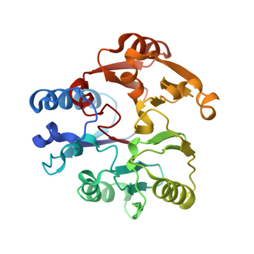
Promiscuous partitioning of a covalent intermediate common in the pentein superfamily.

Linsky, T.W.Monzingo, A.F.Stone, E.M.Robertus, J.D.Fast, W.

(2008) Chem Biol 15: 467-475

  • DOI: https://doi.org/10.1016/j.chembiol.2008.03.012
  • Primary Citation of Related Structures:  
    3BPB

  • PubMed Abstract: 

    Many enzymes in the pentein superfamily use a transient covalent intermediate in their catalytic mechanisms. Here we trap and determine the structure of a stable covalent adduct that mimics this intermediate using a mutant dimethylarginine dimethylaminohydrolase and an alternative substrate. The interactions observed between the enzyme and trapped adduct suggest an altered angle of attack between the nucleophiles of the first and second half-reactions of normal catalysis. The stable covalent adduct is also capable of further reaction. Addition of imidazole rescues the original hydrolytic activity. Notably, addition of other amines instead yields substituted arginine products, which arise from partitioning of the intermediate into the evolutionarily related amidinotransferase reaction pathway. The enzyme provides both selectivity and catalysis for the amidinotransferase reaction, underscoring commonalities among the reaction pathways in this mechanistically diverse enzyme superfamily. The promiscuous partitioning of this intermediate may also help to illuminate the evolutionary history of these enzymes.


  • Organizational Affiliation

    Department of Chemistry and Biochemistry, College of Pharmacy, The University of Texas, Austin, TX 78712, USA.


Macromolecules
Find similar proteins by:  (by identity cutoff)  |  3D Structure
Entity ID: 1
MoleculeChains Sequence LengthOrganismDetailsImage
dimethylarginine dimethylaminohydrolase
A, B
254Pseudomonas aeruginosaMutation(s): 1 
Gene Names: PA1195
EC: 3.5.3.18
UniProt
Find proteins for Q9I4E3 (Pseudomonas aeruginosa (strain ATCC 15692 / DSM 22644 / CIP 104116 / JCM 14847 / LMG 12228 / 1C / PRS 101 / PAO1))
Explore Q9I4E3 
Go to UniProtKB:  Q9I4E3
Entity Groups  
Sequence Clusters30% Identity50% Identity70% Identity90% Identity95% Identity100% Identity
UniProt GroupQ9I4E3
Sequence Annotations
Expand
  • Reference Sequence
Small Molecules
Ligands 1 Unique
IDChains Name / Formula / InChI Key2D Diagram3D Interactions
SMZ
Query on SMZ

Download Ideal Coordinates CCD File 
C [auth A],
D [auth B]
N~5~-[(E)-imino(methylsulfanyl)methyl]-L-ornithine
C7 H15 N3 O2 S
NGVMVBQRKZPFLB-YFKPBYRVSA-N
Experimental Data & Validation

Experimental Data

  • Method: X-RAY DIFFRACTION
  • Resolution: 2.81 Å
  • R-Value Free: 0.297 
  • R-Value Work: 0.233 
  • Space Group: P 21 21 2
Unit Cell:
Length ( Å )Angle ( ˚ )
a = 86.258α = 90
b = 127.532β = 90
c = 47.339γ = 90
Software Package:
Software NamePurpose
DENZOdata reduction
SCALEPACKdata scaling
MOLREPphasing
CNSrefinement
PDB_EXTRACTdata extraction
CrystalCleardata collection
HKL-2000data reduction
HKL-2000data scaling

Structure Validation

View Full Validation Report


Ligand Structure Quality Assessment 


Entry History 

Deposition Data

Revision History  (Full details and data files)

  • Version 1.0: 2008-06-17
    Type: Initial release
  • Version 1.1: 2011-07-13
    Changes: Version format compliance
  • Version 1.2: 2017-10-25
    Changes: Advisory, Refinement description
  • Version 1.3: 2021-10-20
    Changes: Database references, Derived calculations
  • Version 1.4: 2023-08-30
    Changes: Data collection, Refinement description