3BN9

Crystal Structure of MT-SP1 in complex with Fab Inhibitor E2


Experimental Data Snapshot

  • Method: X-RAY DIFFRACTION
  • Resolution: 2.17 Å
  • R-Value Free: 0.267 
  • R-Value Work: 0.223 
  • R-Value Observed: 0.226 

wwPDB Validation   3D Report Full Report


This is version 1.4 of the entry. See complete history


Literature

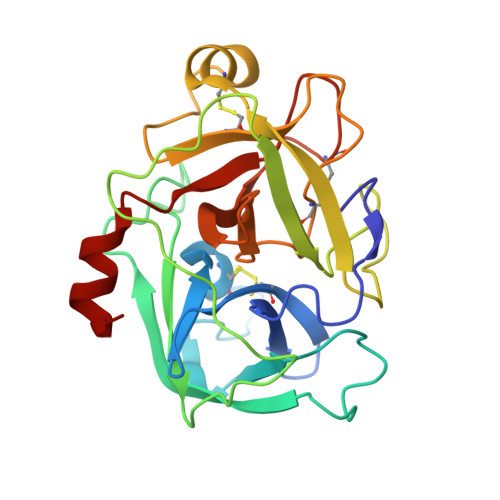
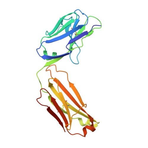
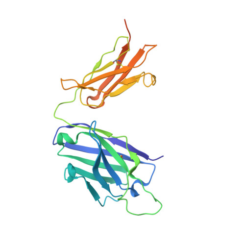
Structure of an Fab-protease complex reveals a highly specific non-canonical mechanism of inhibition

Farady, C.J.Egea, P.F.Schneider, E.L.Darragh, M.R.Craik, C.S.

(2008) J Mol Biol 380: 351-360

  • DOI: https://doi.org/10.1016/j.jmb.2008.05.009
  • Primary Citation of Related Structures:  
    3BN9

  • PubMed Abstract: 

    The vast majority of protein protease inhibitors bind their targets in a substrate-like manner. This is a robust and efficient mechanism of inhibition but, due to the highly conserved architecture of protease active sites, these inhibitors often exhibit promiscuity. Inhibitors that show strict specificity for one protease usually achieve this selectivity by combining substrate-like binding in the active site with exosite binding on the protease surface. The development of new, specific inhibitors can be aided greatly by binding to non-conserved regions of proteases if potency can be maintained. Due to their ability to bind specifically to nearly any antigen, antibodies provide an excellent scaffold for creating inhibitors targeted to a single member of a family of highly homologous enzymes. The 2.2 A resolution crystal structure of an Fab antibody inhibitor in complex with the serine protease membrane-type serine protease 1 (MT-SP1/matriptase) reveals the molecular basis of its picomolar potency and specificity. The inhibitor has a distinct mechanism of inhibition; it gains potency and specificity through interactions with the protease surface loops, and inhibits by binding in the active site in a catalytically non-competent manner. In contrast to most naturally occurring protease inhibitors, which have diverse structures but converge to a similar inhibitory archetype, antibody inhibitors provide an opportunity to develop divergent mechanisms of inhibition from a single scaffold.


  • Organizational Affiliation

    Graduate Group in Biophysics, University of California, San Francisco, 600 16th St. Genentech Hall, San Francisco, CA 94143-2240, USA.


Macromolecules
Find similar proteins by:  (by identity cutoff)  |  3D Structure
Entity ID: 1
MoleculeChains Sequence LengthOrganismDetailsImage
Membrane-type serine protease 1A [auth B],
D [auth A]
241Homo sapiensMutation(s): 1 
Gene Names: ST14PRSS14SNC19TADG15
EC: 3.4.21.109
UniProt & NIH Common Fund Data Resources
Find proteins for Q9Y5Y6 (Homo sapiens)
Explore Q9Y5Y6 
Go to UniProtKB:  Q9Y5Y6
PHAROS:  Q9Y5Y6
GTEx:  ENSG00000149418 
Entity Groups  
Sequence Clusters30% Identity50% Identity70% Identity90% Identity95% Identity100% Identity
UniProt GroupQ9Y5Y6
Sequence Annotations
Expand
  • Reference Sequence
Find similar proteins by:  (by identity cutoff)  |  3D Structure
Entity ID: 2
MoleculeChains Sequence LengthOrganismDetailsImage
E2 Fab Light ChainB [auth C],
E
214Homo sapiensMutation(s): 0 
Entity Groups  
Sequence Clusters30% Identity50% Identity70% Identity90% Identity95% Identity100% Identity
Sequence Annotations
Expand
  • Reference Sequence
Find similar proteins by:  (by identity cutoff)  |  3D Structure
Entity ID: 3
MoleculeChains Sequence LengthOrganismDetailsImage
E2 Fab Heavy ChainC [auth D],
F
257Homo sapiensMutation(s): 0 
UniProt & NIH Common Fund Data Resources
Find proteins for P01764 (Homo sapiens)
Explore P01764 
Go to UniProtKB:  P01764
PHAROS:  P01764
GTEx:  ENSG00000211949 
Entity Groups  
Sequence Clusters30% Identity50% Identity70% Identity90% Identity95% Identity100% Identity
UniProt GroupP01764
Sequence Annotations
Expand
  • Reference Sequence
Small Molecules
Ligands 2 Unique
IDChains Name / Formula / InChI Key2D Diagram3D Interactions
SO4
Query on SO4

Download Ideal Coordinates CCD File 
M [auth C],
T [auth E]
SULFATE ION
O4 S
QAOWNCQODCNURD-UHFFFAOYSA-L
EDO
Query on EDO

Download Ideal Coordinates CCD File 
G [auth B]
H [auth B]
I [auth B]
J [auth B]
K [auth B]
G [auth B],
H [auth B],
I [auth B],
J [auth B],
K [auth B],
L [auth B],
N [auth C],
O [auth C],
P [auth D],
Q [auth A],
R [auth A],
S [auth A],
U [auth E]
1,2-ETHANEDIOL
C2 H6 O2
LYCAIKOWRPUZTN-UHFFFAOYSA-N
Experimental Data & Validation

Experimental Data

  • Method: X-RAY DIFFRACTION
  • Resolution: 2.17 Å
  • R-Value Free: 0.267 
  • R-Value Work: 0.223 
  • R-Value Observed: 0.226 
  • Space Group: P 21 21 21
Unit Cell:
Length ( Å )Angle ( ˚ )
a = 48.625α = 90
b = 163.279β = 90
c = 201.16γ = 90
Software Package:
Software NamePurpose
DENZOdata reduction
SCALEPACKdata scaling
PHASERphasing
PHENIXrefinement
PDB_EXTRACTdata extraction
ELVESdata reduction

Structure Validation

View Full Validation Report


Entry History 

Deposition Data

Revision History  (Full details and data files)

  • Version 1.0: 2008-09-09
    Type: Initial release
  • Version 1.1: 2011-07-13
    Changes: Version format compliance
  • Version 1.2: 2017-10-25
    Changes: Advisory, Refinement description, Source and taxonomy
  • Version 1.3: 2021-10-20
    Changes: Advisory, Database references, Derived calculations
  • Version 1.4: 2023-08-30
    Changes: Data collection, Refinement description