3BKV

X-ray structure of the bacteriophage phiKZ lytic transglycosylase, gp144, in complex with chitotetraose, (NAG)4


Experimental Data Snapshot

  • Method: X-RAY DIFFRACTION
  • Resolution: 2.60 Å
  • R-Value Free: 0.272 
  • R-Value Work: 0.227 
  • R-Value Observed: 0.227 

wwPDB Validation   3D Report Full Report


This is version 2.2 of the entry. See complete history


Literature

Structure of the bacteriophage phi KZ lytic transglycosylase gp144.

Fokine, A.Miroshnikov, K.A.Shneider, M.M.Mesyanzhinov, V.V.Rossmann, M.G.

(2008) J Biol Chem 283: 7242-7250

  • DOI: https://doi.org/10.1074/jbc.M709398200
  • Primary Citation of Related Structures:  
    3BKH, 3BKV

  • PubMed Abstract: 

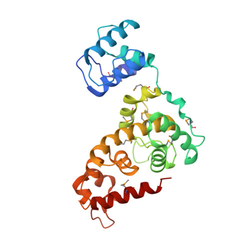
    Lytic transglycosylases are enzymes that act on the peptidoglycan of bacterial cell walls. They cleave the glycosidic linkage between N-acetylmuramoyl and N-acetylglucosaminyl residues with the concomitant formation of a 1,6-anhydromuramoyl product. The x-ray structure of the lytic transglycosylase gp144 from the Pseudomonas bacteriophage phi KZ has been determined to 2.5-A resolution. This protein is probably employed by the bacteriophage in the late stage of the virus reproduction cycle to destroy the bacterial cell wall to release the phage progeny. phi KZ gp144 is a 260-residue alpha-helical protein composed of a 70-residue N-terminal cell wall-binding domain and a C-terminal catalytic domain. The fold of the N-terminal domain is similar to the peptidoglycan-binding domain from Streptomyces albus G D-Ala-D-Ala carboxypeptidase and to the N-terminal prodomain of human metalloproteinases that act on extracellular matrices. The C-terminal catalytic domain of gp144 has a structural similarity to the catalytic domain of the transglycosylase Slt70 from Escherichia coli and to lysozymes. The gp144 catalytic domain has an elongated groove that can bind at least five sugar residues at sites A-E. As in other lysozymes, the peptidoglycan cleavage (catalyzed by Glu 115 in gp144) occurs between sugar-binding subsites D and E. The x-ray structure of the phi KZ transglycosylase complexed with the chitotetraose (N-acetylglucosamine)(4) has been determined to 2.6-A resolution. The N-acetylglucosamine residues of the chitotetraose bind in sites A-D.


  • Organizational Affiliation

    Department of Biological Sciences, Purdue University, West Lafayette, Indiana 47907-2054, USA.


Macromolecules
Find similar proteins by:  (by identity cutoff)  |  3D Structure
Entity ID: 1
MoleculeChains Sequence LengthOrganismDetailsImage
lytic transglycosylase268Phikzvirus phiKZMutation(s): 0 
Gene Names: ORF144
UniProt
Find proteins for Q8SD18 (Pseudomonas phage phiKZ)
Explore Q8SD18 
Go to UniProtKB:  Q8SD18
Entity Groups  
Sequence Clusters30% Identity50% Identity70% Identity90% Identity95% Identity100% Identity
UniProt GroupQ8SD18
Sequence Annotations
Expand
  • Reference Sequence
Oligosaccharides

Help

Entity ID: 2
MoleculeChains Length2D Diagram Glycosylation3D Interactions
2-acetamido-2-deoxy-beta-D-glucopyranose-(1-4)-2-acetamido-2-deoxy-beta-D-glucopyranose-(1-4)-2-acetamido-2-deoxy-beta-D-glucopyranose-(1-4)-2-acetamido-2-deoxy-beta-D-glucopyranose
B
4N/A
Glycosylation Resources
GlyTouCan:  G01361SX
GlyCosmos:  G01361SX
GlyGen:  G01361SX
Small Molecules
Modified Residues  1 Unique
IDChains TypeFormula2D DiagramParent
MSE
Query on MSE
A
L-PEPTIDE LINKINGC5 H11 N O2 SeMET
Experimental Data & Validation

Experimental Data

  • Method: X-RAY DIFFRACTION
  • Resolution: 2.60 Å
  • R-Value Free: 0.272 
  • R-Value Work: 0.227 
  • R-Value Observed: 0.227 
  • Space Group: P 61
Unit Cell:
Length ( Å )Angle ( ˚ )
a = 62.322α = 90
b = 62.322β = 90
c = 182.643γ = 120
Software Package:
Software NamePurpose
HKL-2000data collection
CNSrefinement
HKL-2000data reduction
HKL-2000data scaling
CNSphasing

Structure Validation

View Full Validation Report


Entry History 

Deposition Data

Revision History  (Full details and data files)

  • Version 1.0: 2008-01-08
    Type: Initial release
  • Version 1.1: 2011-07-13
    Changes: Non-polymer description, Version format compliance
  • Version 2.0: 2020-07-29
    Type: Remediation
    Reason: Carbohydrate remediation
    Changes: Atomic model, Data collection, Database references, Derived calculations, Structure summary
  • Version 2.1: 2023-08-30
    Changes: Data collection, Database references, Refinement description, Structure summary
  • Version 2.2: 2023-11-15
    Changes: Data collection