3BKQ | pdb_00003bkq

Structure of the P368G mutant of PMM/PGM in complex with its substrate


Experimental Data Snapshot

  • Method: X-RAY DIFFRACTION
  • Resolution: 2.05 Å
  • R-Value Free: 
    0.249 (Depositor), 0.248 (DCC) 
  • R-Value Work: 
    0.218 (Depositor), 0.218 (DCC) 
  • R-Value Observed: 
    0.220 (Depositor) 

Starting Model: experimental
View more details

wwPDB Validation 3D Report Full Report

Validation slider image for 3BKQ

Ligand Structure Quality Assessment 


This is version 1.5 of the entry. See complete history

Literature

Backbone flexibility, conformational change, and catalysis in a phosphohexomutase from Pseudomonas aeruginosa.

Schramm, A.M.Mehra-Chaudhary, R.Furdui, C.M.Beamer, L.J.

(2008) Biochemistry 47: 9154-9162

  • DOI: https://doi.org/10.1021/bi8005219
  • Primary Citation Related Structures: 
    3BKQ, 3C04

  • PubMed Abstract: 

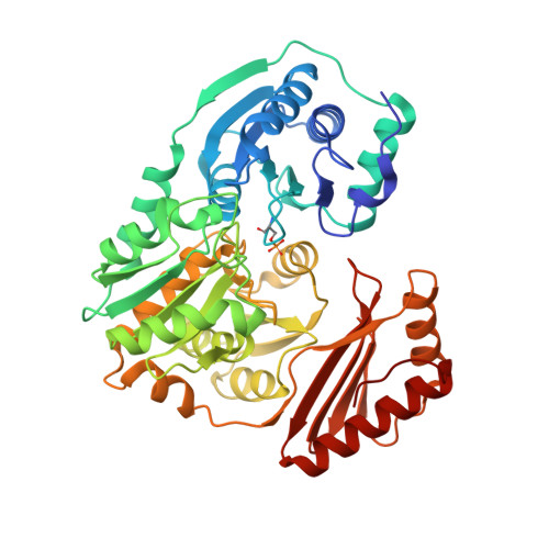
    The enzyme phosphomannomutase/phosphoglucomutase (PMM/PGM) from the bacterium Pseudomonas aeruginosa is involved in the biosynthesis of several complex carbohydrates, including alginate, lipopolysaccharide, and rhamnolipid. Previous structural studies of this protein have shown that binding of substrates produces a rotation of the C-terminal domain, changing the active site from an open cleft in the apoenzyme into a deep, solvent inaccessible pocket where phosphoryl transfer takes place. We report herein site-directed mutagenesis, kinetic, and structural studies in examining the role of residues in the hinge between domains 3 and 4, as well as residues that participate in enzyme-substrate contacts and help form the multidomain "lid" of the active site. We find that the backbone flexibility of residues in the hinge region (e.g., mutation of proline to glycine/alanine) affects the efficiency of the reaction, decreasing k cat by approximately 10-fold and increasing K m by approximately 2-fold. Moreover, thermodynamic analyses show that these changes are due primarily to entropic effects, consistent with an increase in the flexibility of the polypeptide backbone leading to a decreased probability of forming a catalytically productive active site. These results for the hinge residues contrast with those for mutants in the active site of the enzyme, which have profound effects on enzyme kinetics (10 (2)-10 (3)-fold decrease in k cat/ K m) and also show substantial differences in their thermodynamic parameters relative to those of the wild-type (WT) enzyme. These studies support the concept that polypeptide flexibility in protein hinges may evolve to optimize and tune reaction rates.


  • Organizational Affiliation
    • Department of Biochemistry, University of Missouri, Columbia, Missouri 65211, USA.

Macromolecule Content 

  • Total Structure Weight: 50.72 kDa 
  • Atom Count: 3,675 
  • Modeled Residue Count: 458 
  • Deposited Residue Count: 463 
  • Unique protein chains: 1

Macromolecules

Find similar proteins by:|  3D Structure
Entity ID: 1
MoleculeChains  Sequence LengthOrganismDetailsImage
Phosphomannomutase/phosphoglucomutaseA [auth X]463Pseudomonas aeruginosaMutation(s): 1 
Gene Names: algC
EC: 5.4.2.8 (PDB Primary Data), 5.4.2.2 (PDB Primary Data)
UniProt
Find proteins for P26276 (Pseudomonas aeruginosa (strain ATCC 15692 / DSM 22644 / CIP 104116 / JCM 14847 / LMG 12228 / 1C / PRS 101 / PAO1))
Explore P26276 
Go to UniProtKB:  P26276
Entity Groups
Sequence Clusters30% Identity50% Identity70% Identity90% Identity95% Identity100% Identity
UniProt GroupP26276
Sequence Annotations
Expand
Reference Sequence

Small Molecules

Modified Residues  1 Unique
IDChains TypeFormula2D DiagramParent
SEP
Query on SEP
A [auth X]L-PEPTIDE LINKINGC3 H8 N O6 PSER

Experimental Data & Validation

Experimental Data

  • Method: X-RAY DIFFRACTION
  • Resolution: 2.05 Å
  • R-Value Free:  0.249 (Depositor), 0.248 (DCC) 
  • R-Value Work:  0.218 (Depositor), 0.218 (DCC) 
  • R-Value Observed: 0.220 (Depositor) 
Space Group: P 21 21 21
Unit Cell:
Length ( Å )Angle ( ˚ )
a = 70.604α = 90
b = 74.74β = 90
c = 86.924γ = 90
Software Package:
Software NamePurpose
REFMACrefinement
d*TREKdata reduction
d*TREKdata scaling

Structure Validation

View Full Validation Report


Ligand Structure Quality Assessment 


Entry History 

Deposition Data

Revision History  (Full details and data files)

  • Version 1.0: 2008-09-09
    Type: Initial release
  • Version 1.1: 2011-07-13
    Changes: Advisory, Version format compliance
  • Version 1.2: 2020-07-29
    Type: Remediation
    Reason: Carbohydrate remediation
    Changes: Data collection, Derived calculations, Structure summary
  • Version 1.3: 2021-10-20
    Changes: Database references, Structure summary
  • Version 1.4: 2023-08-30
    Changes: Data collection, Refinement description
  • Version 1.5: 2024-11-06
    Changes: Structure summary