3BKK | pdb_00003bkk

Tesis ACE co-crystal structure with ketone ACE inhibitor kAF


Experimental Data Snapshot

  • Method: X-RAY DIFFRACTION
  • Resolution: 2.17 Å
  • R-Value Free: 
    0.251 (Depositor), 0.255 (DCC) 
  • R-Value Work: 
    0.202 (Depositor), 0.207 (DCC) 

Starting Model: experimental
View more details

wwPDB Validation 3D Report Full Report

Validation slider image for 3BKK

Ligand Structure Quality Assessment 


This is version 2.3 of the entry. See complete history

Literature

Probing the basis of domain-dependent inhibition using novel ketone inhibitors of Angiotensin-converting enzyme

Watermeyer, J.M.Kroger, W.L.O'Neill, H.G.Sewell, B.T.Sturrock, E.D.

(2008) Biochemistry 47: 5942-5950

  • DOI: https://doi.org/10.1021/bi8002605
  • Primary Citation Related Structures: 
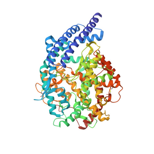
    3BKK, 3BKL

  • PubMed Abstract: 

    Human angiotensin-converting enzyme (ACE) has two homologous domains, the N and C domains, with differing substrate preferences. X-ray crystal structures of the C and N domains complexed with various inhibitors have allowed identification of active site residues that might be important for the molecular basis of this selectivity. However, it is unclear to what extent the different residues contribute to substrate domain selectivity. Here, cocrystal structures of human testis ACE, equivalent to the C domain, have been determined with two novel C domain-selective ketomethylene inhibitors, (5 S)-5-[( N-benzoyl)amino]-4-oxo-6-phenylhexanoyl- l-tryptophan (kAW) and (5 S)-5-[( N-benzoyl)amino]-4-oxo-6-phenylhexanoyl- l-phenylalanine (kAF). The ketone groups of both inhibitors bind to the zinc ion as a hydrated geminal diolate, demonstrating the ability of the active site to catalyze the formation of the transition state. Moreover, active site residues involved in inhibitor binding have been mutated to their N domain counterparts, and the effect of the mutations on inhibitor binding has been determined. The C domain selectivity of these inhibitors was found to result from interactions between bulky hydrophobic side chain moieties and C domain-specific residues F391, V518, E376, and V380 (numbering of testis ACE). Mutation of these residues decreased the affinity for the inhibitors 4-20-fold. T282, V379, E403, D453, and S516 did not contribute individually to C domain-selective inhibitor binding. Further domain-selective inhibitor design should focus on increasing both the affinity and selectivity of the side chain moieties.


  • Organizational Affiliation
    • Division of Medical Biochemistry, Institute of Infectious Disease and Molecular Medicine, University of Cape Town, Observatory 7925, South Africa.

Macromolecule Content 

  • Total Structure Weight: 69.92 kDa 
  • Atom Count: 5,181 
  • Modeled Residue Count: 586 
  • Deposited Residue Count: 591 
  • Unique protein chains: 1

Macromolecules

Find similar proteins by:|  3D Structure
Entity ID: 1
MoleculeChains  Sequence LengthOrganismDetailsImage
Angiotensin-converting enzyme, somatic isoform591Homo sapiensMutation(s): 5 
Gene Names: ACEDCPDCP1
EC: 3.4.15.1
UniProt & NIH Common Fund Data Resources
Find proteins for P12821 (Homo sapiens)
Explore P12821 
Go to UniProtKB:  P12821
PHAROS:  P12821
GTEx:  ENSG00000159640 
Entity Groups
Sequence Clusters30% Identity50% Identity70% Identity90% Identity95% Identity100% Identity
UniProt GroupP12821
Glycosylation
Glycosylation Sites: 2Go to GlyGen: P12821-1
Sequence Annotations
Expand
Reference Sequence

Oligosaccharides

Help  
Entity ID: 2
MoleculeChains Length2D Diagram GlycosylationD Interactions
2-acetamido-2-deoxy-beta-D-glucopyranose-(1-4)-[alpha-L-fucopyranose-(1-6)]2-acetamido-2-deoxy-beta-D-glucopyranose
B
3N-Glycosylation
Glycosylation Resources
GlyTouCan: G21290RB
GlyCosmos: G21290RB
GlyGen: G21290RB

Small Molecules

Ligands 4 Unique
IDChains Name / Formula / InChI Key2D Diagram3D Interactions
KAF

Query on KAF


Download:Ideal Coordinates CCD File
H [auth A]N-{(5S)-4,4-dihydroxy-6-phenyl-5-[(phenylcarbonyl)amino]hexanoyl}-L-phenylalanine
C28 H30 N2 O6
XSYPPQBCHNRTIT-ZEQRLZLVSA-N
NAG

Query on NAG


Download:Ideal Coordinates CCD File
C [auth A],
D [auth A]
2-acetamido-2-deoxy-beta-D-glucopyranose
C8 H15 N O6
OVRNDRQMDRJTHS-FMDGEEDCSA-N
ZN

Query on ZN


Download:Ideal Coordinates CCD File
E [auth A]ZINC ION
Zn
PTFCDOFLOPIGGS-UHFFFAOYSA-N
CL

Query on CL


Download:Ideal Coordinates CCD File
F [auth A],
G [auth A]
CHLORIDE ION
Cl
VEXZGXHMUGYJMC-UHFFFAOYSA-M

Experimental Data & Validation

Experimental Data

  • Method: X-RAY DIFFRACTION
  • Resolution: 2.17 Å
  • R-Value Free:  0.251 (Depositor), 0.255 (DCC) 
  • R-Value Work:  0.202 (Depositor), 0.207 (DCC) 
Space Group: P 21 21 21
Unit Cell:
Length ( Å )Angle ( ˚ )
a = 60.383α = 90
b = 85.07β = 90
c = 135.506γ = 90
Software Package:
Software NamePurpose
DENZOdata reduction
SCALEPACKdata scaling
EPMRphasing
CNSrefinement
PDB_EXTRACTdata extraction
HKL-2000data collection
HKL-2000data reduction
HKL-2000data scaling

Structure Validation

View Full Validation Report


Ligand Structure Quality Assessment 


Entry History 

Deposition Data

Revision History  (Full details and data files)

  • Version 1.0: 2008-06-10
    Type: Initial release
  • Version 1.1: 2011-07-13
    Changes: Non-polymer description, Version format compliance
  • Version 1.2: 2017-10-25
    Changes: Refinement description, Source and taxonomy
  • Version 2.0: 2020-07-29
    Type: Remediation
    Reason: Carbohydrate remediation
    Changes: Atomic model, Data collection, Derived calculations, Structure summary
  • Version 2.1: 2021-10-20
    Changes: Database references, Structure summary
  • Version 2.2: 2023-08-30
    Changes: Data collection, Refinement description
  • Version 2.3: 2024-11-13
    Changes: Structure summary