3BK9

H55A mutant of tryptophan 2,3-dioxygenase from Xanthomonas campestris


Experimental Data Snapshot

  • Method: X-RAY DIFFRACTION
  • Resolution: 2.15 Å
  • R-Value Free: 0.285 
  • R-Value Work: 0.200 
  • R-Value Observed: 0.205 

wwPDB Validation   3D Report Full Report


Ligand Structure Quality Assessment 


This is version 1.3 of the entry. See complete history


Literature

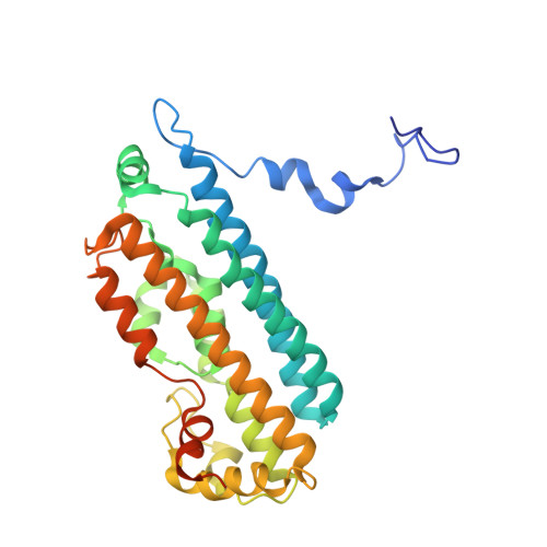
Histidine 55 of tryptophan 2,3-dioxygenase is not an active site base but regulates catalysis by controlling substrate binding

Thackray, S.J.Bruckmann, C.Anderson, J.L.R.Campbell, L.P.Xiao, R.Zhao, L.Mowat, C.G.Forouhar, F.Tong, L.Chapman, S.K.

(2008) Biochemistry 47: 10677-10684

  • DOI: https://doi.org/10.1021/bi801202a
  • Primary Citation of Related Structures:  
    3BK9, 3E08

  • PubMed Abstract: 

    Tryptophan 2,3-dioxygenase (TDO) from Xanthomonas campestris is a highly specific heme-containing enzyme from a small family of homologous enzymes, which includes indoleamine 2,3-dioxygenase (IDO). The structure of wild type (WT TDO) in the catalytically active, ferrous (Fe (2+)) form and in complex with its substrate l-tryptophan ( l-Trp) was recently reported [Forouhar et al. (2007) Proc. Natl. Acad. Sci. U.S.A. 104, 473-478] and revealed that histidine 55 hydrogen bonds to l-Trp, precisely positioning it in the active site and implicating it as a possible active site base. In this study the substitution of the active site residue histidine 55 by alanine and serine (H55A and H55S) provides insight into the molecular mechanism used by the enzyme to control substrate binding. We report the crystal structure of the H55A and H55S mutant forms at 2.15 and 1.90 A resolution, respectively, in binary complexes with l-Trp. These structural data, in conjunction with potentiometric and kinetic studies on both mutants, reveal that histidine 55 is not essential for turnover but greatly disfavors the mechanistically unproductive binding of l-Trp to the oxidized enzyme allowing control of catalysis. This is demonstrated by the difference in the K d values for l-Trp binding to the two oxidation states of wild-type TDO (3.8 mM oxidized, 4.1 microM reduced), H55A TDO (11.8 microM oxidized, 3.7 microM reduced), and H55S TDO (18.4 microM oxidized, 5.3 microM reduced).


  • Organizational Affiliation

    EaStCHEM, School of Chemistry, University of Edinburgh, UK.


Macromolecules
Find similar proteins by:  (by identity cutoff)  |  3D Structure
Entity ID: 1
MoleculeChains Sequence LengthOrganismDetailsImage
Tryptophan 2,3-dioxygenase
A, B, C, D, E
A, B, C, D, E, F, G, H
306Xanthomonas campestris pv. campestrisMutation(s): 1 
EC: 1.13.11.11
UniProt
Find proteins for Q8PDA8 (Xanthomonas campestris pv. campestris (strain ATCC 33913 / DSM 3586 / NCPPB 528 / LMG 568 / P 25))
Explore Q8PDA8 
Go to UniProtKB:  Q8PDA8
Entity Groups  
Sequence Clusters30% Identity50% Identity70% Identity90% Identity95% Identity100% Identity
UniProt GroupQ8PDA8
Sequence Annotations
Expand
  • Reference Sequence
Small Molecules
Ligands 2 Unique
IDChains Name / Formula / InChI Key2D Diagram3D Interactions
HEM
Query on HEM

Download Ideal Coordinates CCD File 
AA [auth G]
CA [auth H]
J [auth A]
M [auth B]
Q [auth C]
AA [auth G],
CA [auth H],
J [auth A],
M [auth B],
Q [auth C],
T [auth D],
V [auth E],
Y [auth F]
PROTOPORPHYRIN IX CONTAINING FE
C34 H32 Fe N4 O4
KABFMIBPWCXCRK-RGGAHWMASA-L
TRP
Query on TRP

Download Ideal Coordinates CCD File 
BA [auth H]
I [auth A]
K [auth A]
L [auth B]
N [auth B]
BA [auth H],
I [auth A],
K [auth A],
L [auth B],
N [auth B],
O [auth C],
P [auth C],
R [auth D],
S [auth D],
U [auth E],
W [auth E],
X [auth F],
Z [auth G]
TRYPTOPHAN
C11 H12 N2 O2
QIVBCDIJIAJPQS-VIFPVBQESA-N
Experimental Data & Validation

Experimental Data

  • Method: X-RAY DIFFRACTION
  • Resolution: 2.15 Å
  • R-Value Free: 0.285 
  • R-Value Work: 0.200 
  • R-Value Observed: 0.205 
  • Space Group: P 1 21 1
Unit Cell:
Length ( Å )Angle ( ˚ )
a = 78.222α = 90
b = 117.606β = 95.73
c = 139.283γ = 90
Software Package:
Software NamePurpose
REFMACrefinement
MOSFLMdata reduction
SCALAdata scaling
AMoREphasing

Structure Validation

View Full Validation Report


Ligand Structure Quality Assessment 


Entry History 

Deposition Data

Revision History  (Full details and data files)

  • Version 1.0: 2008-09-30
    Type: Initial release
  • Version 1.1: 2011-07-13
    Changes: Version format compliance
  • Version 1.2: 2021-11-10
    Changes: Database references, Derived calculations
  • Version 1.3: 2023-11-01
    Changes: Data collection, Refinement description