3BGQ | pdb_00003bgq

Human Pim-1 kinase in complex with an triazolo pyridazine inhibitor VX2


Experimental Data Snapshot

  • Method: X-RAY DIFFRACTION
  • Resolution: 2.00 Å
  • R-Value Free: 
    0.230 (Depositor), 0.217 (DCC) 
  • R-Value Work: 
    0.212 (Depositor), 0.201 (DCC) 
  • R-Value Observed: 
    0.213 (Depositor) 

wwPDB Validation 3D Report Full Report

Validation slider image for 3BGQ

Ligand Structure Quality Assessment 


This is version 1.2 of the entry. See complete history

Literature

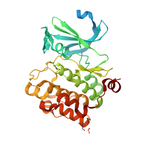
Docking study yields four novel inhibitors of the protooncogene pim-1 kinase.

Pierce, A.C.Jacobs, M.Stuver-Moody, C.

(2008) J Med Chem 51: 1972-1975

  • DOI: https://doi.org/10.1021/jm701248t
  • Primary Citation Related Structures: 
    3BGP, 3BGQ, 3BGZ

  • PubMed Abstract: 

    To supplement the hits from a high throughput screen, docking was performed against Pim-1 kinase. Glide docking was augmented with a filter to require traditional or aromatic CH..O hydrogen bonds to the kinase hinge. Four diverse actives, of 96 molecules assayed, had K(i) values between 0.091 and 4.5 microM. This gives a 14-fold enrichment over the earlier HTS run, and the two crystal structures solved confirmed the binding modes predicted by docking.


  • Organizational Affiliation
    • Vertex Pharmaceuticals Incorporated, 130 Waverly Street, Cambridge, MA 02139, USA. al_pierce@vrtx.com

Macromolecule Content 

  • Total Structure Weight: 38.35 kDa 
  • Atom Count: 2,359 
  • Modeled Residue Count: 273 
  • Deposited Residue Count: 333 
  • Unique protein chains: 1

Macromolecules

Find similar proteins by:|  3D Structure
Entity ID: 1
MoleculeChains  Sequence LengthOrganismDetailsImage
Proto-oncogene serine/threonine-protein kinase Pim-1333Homo sapiensMutation(s): 0 
Gene Names: PIM1
EC: 2.7.11.1
UniProt & NIH Common Fund Data Resources
Find proteins for P11309 (Homo sapiens)
Explore P11309 
Go to UniProtKB:  P11309
PHAROS:  P11309
GTEx:  ENSG00000137193 
Entity Groups
Sequence Clusters30% Identity50% Identity70% Identity90% Identity95% Identity100% Identity
UniProt GroupP11309
Sequence Annotations
Expand
Reference Sequence

Small Molecules

Ligands 1 Unique
IDChains Name / Formula / InChI Key2D Diagram3D Interactions
VX2

Query on VX2


Download:Ideal Coordinates CCD File
B [auth A]N-cyclohexyl-3-[3-(trifluoromethyl)phenyl][1,2,4]triazolo[4,3-b]pyridazin-6-amine
C18 H18 F3 N5
XYYDXQCAYXOGQT-UHFFFAOYSA-N
Modified Residues  1 Unique
IDChains TypeFormula2D DiagramParent
SEP
Query on SEP
A
L-PEPTIDE LINKINGC3 H8 N O6 PSER

Experimental Data & Validation

Experimental Data

  • Method: X-RAY DIFFRACTION
  • Resolution: 2.00 Å
  • R-Value Free:  0.230 (Depositor), 0.217 (DCC) 
  • R-Value Work:  0.212 (Depositor), 0.201 (DCC) 
  • R-Value Observed: 0.213 (Depositor) 
Space Group: P 65
Unit Cell:
Length ( Å )Angle ( ˚ )
a = 98.34α = 90
b = 98.34β = 90
c = 81.35γ = 120
Software Package:
Software NamePurpose
d*TREKdata scaling
CNSrefinement
PDB_EXTRACTdata extraction
d*TREKdata reduction
CNXrefinement

Structure Validation

View Full Validation Report


Ligand Structure Quality Assessment 


Entry History 

Deposition Data

  • Released Date: 2007-12-11 
  • Deposition Author(s): Jacobs, M.D.

Revision History  (Full details and data files)

  • Version 1.0: 2007-12-11
    Type: Initial release
  • Version 1.1: 2011-07-13
    Changes: Version format compliance
  • Version 1.2: 2024-10-30
    Changes: Data collection, Database references, Derived calculations, Structure summary