Staphylococcus aureus DsbA Does Not Have a Destabilizing Disulfide: A NEW PARADIGM FOR BACTERIAL OXIDATIVE FOLDING
Heras, B., Kurz, M., Jarrott, R., Shouldice, S.R., Frei, P., Robin, G., Cemazar, M., Thony-Meyer, L., Glockshuber, R., Martin, J.L.(2008) J Biol Chem 283: 4261-4271
- PubMed: 18077463 Search on PubMed
- DOI: https://doi.org/10.1074/jbc.M707838200
- Primary Citation Related Structures:
3BCI, 3BCK, 3BD2 - PubMed Abstract:
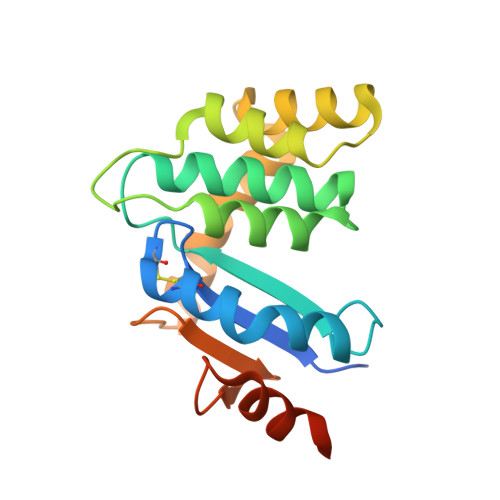
In Gram-negative bacteria, the introduction of disulfide bonds into folding proteins occurs in the periplasm and is catalyzed by donation of an energetically unstable disulfide from DsbA, which is subsequently re-oxidized through interaction with DsbB. Gram-positive bacteria lack a classic periplasm but nonetheless encode Dsb-like proteins. Staphylococcus aureus encodes just one Dsb protein, a DsbA, and no DsbB. Here we report the crystal structure of S. aureus DsbA (SaDsbA), which incorporates a thioredoxin fold with an inserted helical domain, like its Escherichia coli counterpart EcDsbA, but it lacks the characteristic hydrophobic patch and has a truncated binding groove near the active site. These findings suggest that SaDsbA has a different substrate specificity than EcDsbA. Thermodynamic studies indicate that the oxidized and reduced forms of SaDsbA are energetically equivalent, in contrast to the energetically unstable disulfide form of EcDsbA. Further, the partial complementation of EcDsbA by SaDsbA is independent of EcDsbB and biochemical assays show that SaDsbA does not interact with EcDsbB. The identical stabilities of oxidized and reduced SaDsbA may facilitate direct re-oxidation of the protein by extracellular oxidants, without the need for DsbB.
- Institute for Molecular Bioscience, University of Queensland, Brisbane QLD 4072, Australia. bheras@imb.uq.edu.au
Organizational Affiliation: