3BCH | pdb_00003bch

Crystal Structure of the Human Laminin Receptor Precursor


Experimental Data Snapshot

  • Method: X-RAY DIFFRACTION
  • Resolution: 2.15 Å
  • R-Value Free: 
    0.224 (Depositor), 0.224 (DCC) 
  • R-Value Work: 
    0.185 (Depositor), 0.186 (DCC) 
  • R-Value Observed: 
    0.187 (Depositor) 

wwPDB Validation 3D Report Full Report

Validation slider image for 3BCH

This is version 1.2 of the entry. See complete history

Literature

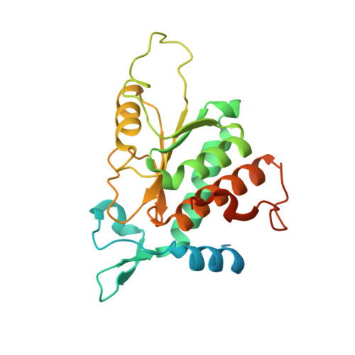
Crystal structure of the human laminin receptor precursor.

Jamieson, K.V.Wu, J.Hubbard, S.R.Meruelo, D.

(2008) J Biological Chem 283: 3002-3005

  • DOI: https://doi.org/10.1074/jbc.C700206200
  • Primary Citation Related Structures: 
    3BCH

  • PubMed Abstract: 

    The human laminin receptor (LamR) interacts with many ligands, including laminin, prions, Sindbis virus, and the polyphenol (-)-epigallocatechin-3-gallate (EGCG), and has been implicated in a number of diseases. LamR is overexpressed on tumor cells, and targeting LamR elicits anti-cancer effects. Here, we report the crystal structure of human LamR, which provides insights into its function and should facilitate the design of novel therapeutics targeting LamR.


  • Organizational Affiliation
    • Gene Therapy Center, Cancer Institute and Department of Pathology, New York University School of Medicine, New York, New York 10016.

Macromolecule Content 

  • Total Structure Weight: 28.21 kDa 
  • Atom Count: 1,710 
  • Modeled Residue Count: 197 
  • Deposited Residue Count: 253 
  • Unique protein chains: 1

Macromolecules

Find similar proteins by:|  3D Structure
Entity ID: 1
MoleculeChains  Sequence LengthOrganismDetailsImage
40S ribosomal protein SA253Homo sapiensMutation(s): 0 
Gene Names: RPSALAMBRLAMR1
UniProt & NIH Common Fund Data Resources
Find proteins for P08865 (Homo sapiens)
Explore P08865 
Go to UniProtKB:  P08865
PHAROS:  P08865
GTEx:  ENSG00000168028 
Entity Groups
Sequence Clusters30% Identity50% Identity70% Identity90% Identity95% Identity100% Identity
UniProt GroupP08865
Sequence Annotations
Expand
Reference Sequence

Experimental Data & Validation

Experimental Data

  • Method: X-RAY DIFFRACTION
  • Resolution: 2.15 Å
  • R-Value Free:  0.224 (Depositor), 0.224 (DCC) 
  • R-Value Work:  0.185 (Depositor), 0.186 (DCC) 
  • R-Value Observed: 0.187 (Depositor) 
Space Group: P 43 21 2
Unit Cell:
Length ( Å )Angle ( ˚ )
a = 75.688α = 90
b = 75.688β = 90
c = 99.007γ = 90
Software Package:
Software NamePurpose
REFMACrefinement

Structure Validation

View Full Validation Report


Entry History 

Deposition Data

Revision History  (Full details and data files)

  • Version 1.0: 2007-12-04
    Type: Initial release
  • Version 1.1: 2011-07-13
    Changes: Version format compliance
  • Version 1.2: 2024-02-21
    Changes: Data collection, Database references