3BAP

Crystal Structure of the 25 kDa Subunit of Human Cleavage Factor Im


Experimental Data Snapshot

  • Method: X-RAY DIFFRACTION
  • Resolution: 1.85 Å
  • R-Value Free: 0.244 
  • R-Value Work: 0.226 
  • R-Value Observed: 0.226 

wwPDB Validation   3D Report Full Report


This is version 1.2 of the entry. See complete history


Literature

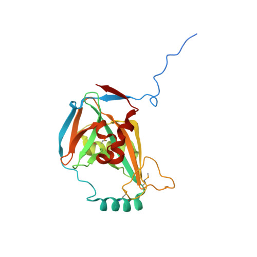
Crystal structure of the 25 kDa subunit of human cleavage factor Im.

Coseno, M.Martin, G.Berger, C.Gilmartin, G.Keller, W.Doublie, S.

(2008) Nucleic Acids Res 36: 3474-3483

  • DOI: https://doi.org/10.1093/nar/gkn079
  • Primary Citation of Related Structures:  
    3BAP, 3BHO

  • PubMed Abstract: 

    Cleavage factor I(m) is an essential component of the pre-messenger RNA 3'-end processing machinery in higher eukaryotes, participating in both the polyadenylation and cleavage steps. Cleavage factor I(m) is an oligomer composed of a small 25 kDa subunit (CF I(m)25) and a variable larger subunit of either 59, 68 or 72 kDa. The small subunit also interacts with RNA, poly(A) polymerase, and the nuclear poly(A)-binding protein. These protein-protein interactions are thought to be facilitated by the Nudix domain of CF I(m)25, a hydrolase motif with a characteristic alpha/beta/alpha fold and a conserved catalytic sequence or Nudix box. We present here the crystal structures of human CF I(m)25 in its free and diadenosine tetraphosphate (Ap(4)A) bound forms at 1.85 and 1.80 A, respectively. CF I(m)25 crystallizes as a dimer and presents the classical Nudix fold. Results from crystallographic and biochemical experiments suggest that CF I(m)25 makes use of its Nudix fold to bind but not hydrolyze ATP and Ap(4)A. The complex and apo protein structures provide insight into the active oligomeric state of CF I(m) and suggest a possible role of nucleotide binding in either the polyadenylation and/or cleavage steps of pre-messenger RNA 3'-end processing.


  • Organizational Affiliation

    Department of Microbiology and Department of Molecular Genetics, University of Vermont, Burlington, VT 05405, USA.


Macromolecules
Find similar proteins by:  (by identity cutoff)  |  3D Structure
Entity ID: 1
MoleculeChains Sequence LengthOrganismDetailsImage
Cleavage and polyadenylation specificity factor subunit 5227Homo sapiensMutation(s): 0 
Gene Names: NUDT21CFIM25CPSF25CPSF5
UniProt & NIH Common Fund Data Resources
Find proteins for O43809 (Homo sapiens)
Explore O43809 
Go to UniProtKB:  O43809
PHAROS:  O43809
GTEx:  ENSG00000167005 
Entity Groups  
Sequence Clusters30% Identity50% Identity70% Identity90% Identity95% Identity100% Identity
UniProt GroupO43809
Sequence Annotations
Expand
  • Reference Sequence
Small Molecules
Ligands 1 Unique
IDChains Name / Formula / InChI Key2D Diagram3D Interactions
GOL
Query on GOL

Download Ideal Coordinates CCD File 
B [auth A]GLYCEROL
C3 H8 O3
PEDCQBHIVMGVHV-UHFFFAOYSA-N
Modified Residues  1 Unique
IDChains TypeFormula2D DiagramParent
MSE
Query on MSE
A
L-PEPTIDE LINKINGC5 H11 N O2 SeMET
Experimental Data & Validation

Experimental Data

  • Method: X-RAY DIFFRACTION
  • Resolution: 1.85 Å
  • R-Value Free: 0.244 
  • R-Value Work: 0.226 
  • R-Value Observed: 0.226 
  • Space Group: P 31 2 1
Unit Cell:
Length ( Å )Angle ( ˚ )
a = 80.115α = 90
b = 80.115β = 90
c = 72.209γ = 120
Software Package:
Software NamePurpose
CNSrefinement
PDB_EXTRACTdata extraction
HKL-2000data collection
HKL-2000data reduction
HKL-2000data scaling
CNSphasing

Structure Validation

View Full Validation Report


Entry History 

Deposition Data

Revision History  (Full details and data files)

  • Version 1.0: 2008-02-19
    Type: Initial release
  • Version 1.1: 2011-07-13
    Changes: Non-polymer description, Version format compliance
  • Version 1.2: 2018-01-24
    Changes: Structure summary