


2.1 A Crystal structure of RNase PH from Mycobacterium tuberculosis.
Antczak, A.J., Lekin, T., Segelke, B.W., Berger, J.M.To be published.
Experimental Data Snapshot
Starting Model: experimental
View more details
wwPDB Validation 3D Report Full Report

Entity ID: 1 | |||||
|---|---|---|---|---|---|
| Molecule | Chains | Sequence Length | Organism | Details | Image |
| Ribonuclease PH | 262 | Mycobacterium tuberculosis H37Rv | Mutation(s): 0 Gene Names: rph, rphA, Rv1340, MT1381, MTCY130.25, MTCY02B10.04 EC: 2.7.7.56 | 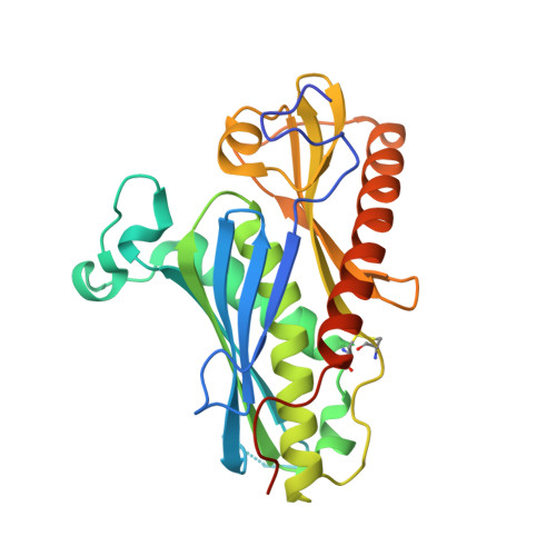 | |
UniProt | |||||
Entity Groups | |||||
| Sequence Clusters | 30% Identity50% Identity70% Identity90% Identity95% Identity100% Identity | ||||
| UniProt Group | P9WGZ7 | ||||
Sequence AnnotationsExpand | |||||
Reference Sequence | |||||
| Ligands 1 Unique | |||||
|---|---|---|---|---|---|
| ID | Chains | Name / Formula / InChI Key | 2D Diagram | 3D Interactions | |
| PO4 Download:Ideal Coordinates CCD File | G [auth A] H [auth A] I [auth B] J [auth C] K [auth C] | PHOSPHATE ION O4 P NBIIXXVUZAFLBC-UHFFFAOYSA-K |  | ||
| Length ( Å ) | Angle ( ˚ ) |
|---|---|
| a = 73.529 | α = 90 |
| b = 152.406 | β = 109.46 |
| c = 83.512 | γ = 90 |
| Software Name | Purpose |
|---|---|
| PHENIX | refinement |
| HKL-2000 | data reduction |
| HKL-2000 | data scaling |
| DM | phasing |


