Minimal pharmacophoric elements and fragment hopping, an approach directed at molecular diversity and isozyme selectivity. Design of selective neuronal nitric oxide synthase inhibitors.
Ji, H., Stanton, B.Z., Igarashi, J., Li, H., Martasek, P., Roman, L.J., Poulos, T.L., Silverman, R.B.(2008) J Am Chem Soc 130: 3900-3914
- PubMed: 18321097 Search on PubMedSearch on PubMed Central
- DOI: https://doi.org/10.1021/ja0772041
- Primary Citation Related Structures:
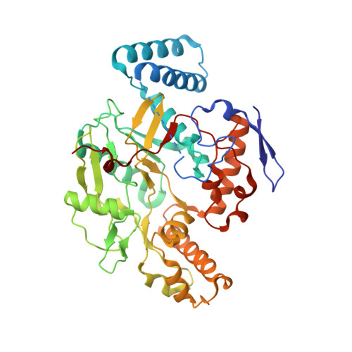
3B3M, 3B3N - PubMed Abstract:
Fragment hopping, a new fragment-based approach for de novo inhibitor design focusing on ligand diversity and isozyme selectivity, is described. The core of this approach is the derivation of the minimal pharmacophoric element for each pharmacophore. Sites for both ligand binding and isozyme selectivity are considered in deriving the minimal pharmacophoric elements. Five general-purpose libraries are established: the basic fragment library, the bioisostere library, the rules for metabolic stability, the toxicophore library, and the side chain library. These libraries are employed to generate focused fragment libraries to match the minimal pharmacophoric elements for each pharmacophore and then to link the fragment to the desired molecule. This method was successfully applied to neuronal nitric oxide synthase (nNOS), which is implicated in stroke and neurodegenerative diseases. Starting with the nitroarginine-containing dipeptide inhibitors we developed previously, a small organic molecule with a totally different chemical structure was designed, which showed nanomolar nNOS inhibitory potency and more than 1000-fold nNOS selectivity. The crystallographic analysis confirms that the small organic molecule with a constrained conformation can exactly mimic the mode of action of the dipeptide nNOS inhibitors. Therefore, a new peptidomimetic strategy, referred to as fragment hopping, which creates small organic molecules that mimic the biological function of peptides by a pharmacophore-driven strategy for fragment-based de novo design, has been established as a new type of fragment-based inhibitor design. As an open system, the newly established approach efficiently incorporates the concept of early "ADME/Tox" considerations and provides a basic platform for medicinal chemistry-driven efforts.
- Department of Chemistry, Center for Drug Discovery and Chemical Biology, Northwestern University, Evanston, Illinois 60208-3113, USA.
Organizational Affiliation: