3B3A | pdb_00003b3a

Structure of E163K/R145E DJ-1


Experimental Data Snapshot

  • Method: X-RAY DIFFRACTION
  • Resolution: 1.50 Å
  • R-Value Free: 
    0.203 (Depositor), 0.198 (DCC) 
  • R-Value Work: 
    0.182 (Depositor), 0.176 (DCC) 
  • R-Value Observed: 
    0.183 (Depositor) 

Starting Model: experimental
View more details

wwPDB Validation 3D Report Full Report

Validation slider image for 3B3A

This is version 1.3 of the entry. See complete history

Literature

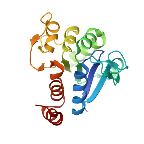
Structural Impact of Three Parkinsonism-Associated Missense Mutations on Human DJ-1.

Lakshminarasimhan, M.Maldonado, M.T.Zhou, W.Fink, A.L.Wilson, M.A.

(2008) Biochemistry 47: 1381-1392

  • DOI: https://doi.org/10.1021/bi701189c
  • Primary Citation Related Structures: 
    2RK3, 2RK4, 2RK6, 3B36, 3B38, 3B3A

  • PubMed Abstract: 

    A number of missense mutations in the oxidative stress response protein DJ-1 are implicated in rare forms of familial Parkinsonism. The best-characterized Parkinsonian DJ-1 missense mutation, L166P, disrupts homodimerization and results in a poorly folded protein. The molecular basis by which the other Parkinsonism-associated mutations disrupt the function of DJ-1, however, is incompletely understood. In this study we show that three different Parkinsonism-associated DJ-1 missense mutations (A104T, E163K, and M26I) reduce the thermal stability of DJ-1 in solution by subtly perturbing the structure of DJ-1 without causing major folding defects or loss of dimerization. Atomic resolution X-ray crystallography shows that the A104T substitution introduces water and a discretely disordered residue into the core of the protein, E163K disrupts a key salt bridge with R145, and M26I causes packing defects in the core of the dimer. The deleterious effect of each Parkinsonism-associated mutation on DJ-1 is dissected by analysis of engineered substitutions (M26L, A104V, and E163K/R145E) that partially alleviate each of the defects introduced by the A104T, E163K and M26I mutations. In total, our results suggest that the protective function of DJ-1 can be compromised by diverse perturbations in its structural integrity, particularly near the junctions of secondary structural elements.


  • Organizational Affiliation
    • Department of Biochemistry and the Redox Biology Center, The University of Nebraska-Lincoln, Lincoln, Nebraska 68588-0664, USA.

Macromolecule Content 

  • Total Structure Weight: 20.39 kDa 
  • Atom Count: 1,630 
  • Modeled Residue Count: 187 
  • Deposited Residue Count: 192 
  • Unique protein chains: 1

Macromolecules

Find similar proteins by:|  3D Structure
Entity ID: 1
MoleculeChains  Sequence LengthOrganismDetailsImage
Protein DJ-1192Homo sapiensMutation(s): 2 
Gene Names: PARK7
EC: 3.5.1 (UniProt), 3.1.2 (UniProt), 3.5.1.124 (UniProt)
UniProt & NIH Common Fund Data Resources
Find proteins for Q99497 (Homo sapiens)
Explore Q99497 
Go to UniProtKB:  Q99497
PHAROS:  Q99497
GTEx:  ENSG00000116288 
Entity Groups
Sequence Clusters30% Identity50% Identity70% Identity90% Identity95% Identity100% Identity
UniProt GroupQ99497
Sequence Annotations
Expand
Reference Sequence

Experimental Data & Validation

Experimental Data

  • Method: X-RAY DIFFRACTION
  • Resolution: 1.50 Å
  • R-Value Free:  0.203 (Depositor), 0.198 (DCC) 
  • R-Value Work:  0.182 (Depositor), 0.176 (DCC) 
  • R-Value Observed: 0.183 (Depositor) 
Space Group: P 31 2 1
Unit Cell:
Length ( Å )Angle ( ˚ )
a = 74.987α = 90
b = 74.987β = 90
c = 75.11γ = 120
Software Package:
Software NamePurpose
REFMACrefinement
CrystalCleardata collection
HKL-2000data reduction
HKL-2000data scaling
PHASERphasing

Structure Validation

View Full Validation Report


Entry History 

Deposition Data

Revision History  (Full details and data files)

  • Version 1.0: 2008-01-15
    Type: Initial release
  • Version 1.1: 2011-07-13
    Changes: Version format compliance
  • Version 1.2: 2021-10-20
    Changes: Database references, Derived calculations
  • Version 1.3: 2023-08-30
    Changes: Data collection, Refinement description